Case Report
Aesthetic protective coverage on molars with MIH: Case report
1 Post-Doctoral Researcher at School of Dentistry of Ribeirão Preto, University of São Paulo (USP)–Ribeirão Preto, São Paulo, Brazil and Professor of Pediatric Clinic and Patients with Disabilities at the Catholic University of Brasilia and IESB University Center, Brasilia, Brazil
2 CEO from Fenelon, Dental Radiographic Diagnosis, Brasília, Brazil
3 CEO from Fenelon, Dental Radiographic Diagnosis, Brasília, Brazil
4 Department of Pediatric Clinics, School of Dentistry of Ribeirão Preto, University of São Paulo (USP)–Ribeirão Preto, São Paulo, Brazil
5 Department of Morphology, Genetics, Orthodontics and Pediatric Dentistry, Araraquara School of Dentistry, São Paulo State University (Unesp), Araraquara, São Paulo, Brazil
Address correspondence to:
Claudia Maria de Souza Peruch
SQS 116 Bloco C, apto 108, Brasilia, CEP 70386030, DF,
Brazil
Access full text article on other devices

Access PDF of article on other devices
Article ID: 100047Z07CP2024
doi: 10.5348/100047Z07CP2024CR
How to cite this article
Peruch CMS, Barriviera M, Barriviera FA, Borsatto MC, Santos-Pinto L. Aesthetic protective coverage on molars with MIH: Case report. J Case Rep Images Dent 2024;10(2):7–13.ABSTRACT
Introduction: The management of molars affected with severe molar incisor hypomineralization (MIH) in young children has been one of the greatest challenges for the pediatric dentist. Determining the severity of the defect, behavior, patient acceptance, and parental expectations are determining factors in defining treatment strategies.
Case Report: The objective of this case report is to demonstrate an aesthetic alternative for preventing fractures of severely affected MIH teeth, using a temporary protective covering. This coverage was carried out using composite resin and a semi-direct technique. The patient’s 9-month follow-up showed favorable adaptation and occlusion and satisfaction from the patient and parents, who placed aesthetics as the main factor for treatment decision.
Conclusion: In this patient, the aesthetic factor was decisive for MIH treatment decision. It was performed an aesthetic protective coverage (APC) using an indirect composite technique.
Introduction
Molar and incisor hypomineralization (MIH) is defined as a qualitative enamel defect that affects one to four first permanent molars and may or may not involve the permanent incisors [1]. It can be clinically observed as a demarcated opacity in the enamel, with a color ranging from creamy white to yellowish-brown. This opaque spot has well-defined edges that identify the limits between the affected enamel and the adjacent healthy enamel [1],[2].
It is estimated that the worldwide prevalence of MIH is 13.5%, making it a public health problem. As important as having this prevalence data is knowing the consequences of this defect for the dentition of the affected patient and so that public health programs can be developed and implemented [3].
Soon after appearing in the mouth, teeth affected by MIH may present painful symptoms, fractures, or caries lesions that progress to the severe destruction of some teeth [3],[4],[5]. Early diagnosis and appropriate therapy can prevent serious complications and improve the patient’s masticatory function and aesthetics [4].
For appropriate therapy, it is important to identify early the affected enamel, the presence of hypersensitivity and the risks of post-eruptive fracture and development of caries lesions. Determining the severity of the MIH defect, the behavior and acceptance of the patient and parents are determining factors for defining treatment strategies [5].
Molar and incisor hypomineralization defects can be considered mild when there are demarcated opacities without post-eruptive enamel fractures, located in areas of little masticatory effort, without sensitivity and absence of caries disease. The MIH defect is considered of moderate severity when the demarcated opacities and post-eruptive fractures affect only the enamel and do not involve cusps and there is no dentin hypersensitivity. However, in severe defects, the demarcated area presents post-eruptive fracture or fracture of part of the dental crown, in addition to being associated with caries disease and presence of dental hypersensitivity [5],[6].
The management of molars affected by severe MIH in young children has been one of the greatest challenges for the pediatric dentist, due to the child’s active growth phase, lack of defined occlusal contacts, wide pulp chambers with prominent pulp horns, and often uncooperative behavior for long sessions. Management may be further aggravated by factors such as porous enamel and rapid disintegration under masticatory load, dental malocclusion, inadequate nutrition of the child, extreme sensitivity and ineffective anesthesia as a result of pulp that presents chronic subclinical inflammation [7]. In addition, frequent failure of restorations due to adhesion problems, associated with secondary caries lesions and successive breaks in hypomineralized enamel results in numerous retreatments with high costs.
The objective of this study is to demonstrate the case of a child with first molars affected by severe MIH in which early diagnosis and temporary aesthetic protective coverage were performed with the aim of preventing fracture of the affected enamel.
Case Report
Patient MCLB, 7 years and 8 months old, came to the office for a routine consultation. The patient was clinically healthy and had no history of systemic problems.
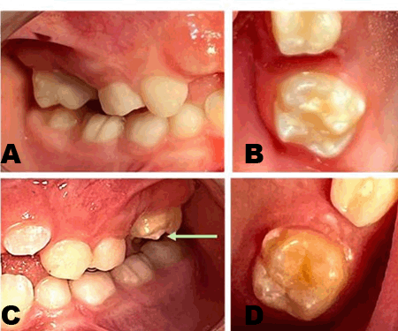
A preventive approach was carried out with education and health promotion through guidance on tooth brushing and correct use of dental floss. After dental prophylaxis, demarcated cream-colored opacities were observed on the vestibular surface and white on the cusps of the first permanent upper molars, diagnosed as severe MIH due to the characteristics of color, location, extension, and a small fracture on the mesial cusp of tooth 26 (Figure 1A, Figure 1B, Figure 1C, Figure 1D).
The parents were concerned about the prognosis of these teeth because the child had been reporting that after eating hard foods, such as popcorn and peanuts, he felt like the tooth was breaking, a fact confirmed in the clinical examination, in which tooth 26, still semi-erupted, showed the beginning of a fracture on the mesiobuccal cusp in the demarcated cream-colored MIH opacity (Figure 1C).
The parents discussed the possibilities of treatment to prevent future fractures. Considering the child’s cooperation with dental procedures, eating habits, occlusion development, and the parents’ anxiety, who expected a temporary aesthetic solution.
We decided to perform a temporary restoration with full coverage, without any preparation, on teeth 16 and 26, which we call aesthetic protective coverage (APC).
To perform the APC, the radiology documentation center (FENELON, Brasília, DF) was asked to provide the digital flow of the patient’s upper and lower arches. After obtaining the digital images of the arches, a “digital wax-up” of 1–1.5 mm thick was performed on teeth 16 and 26 using the NemoStudio software (NemoTech) (Figure 2A, Figure 2B, Figure 2C).
The digital mapping of the initial arch, as well as the arch with the teeth with the virtual wax-up were sent for printing on the Carestream CS 3600 device (CS 3600, Carestream Rochester, NY, USA) (Figure 3A, Figure 3B).
The original digital model and the model with the “waxed” molars were first molded with ZETALABOR dense condensation silicone and finished with ORANWASH light condensation silicone (ZHERMACK). The two printed models and the silicone molding of the wall type for the model, made by the documentation center (FENELON, Brasília, DF) were sent to the pediatric dentist’s office, who chose to make the protective aesthetic coverings in the office itself using the semi-direct technique instead of 3D printing for provisional that could have been done by the documentation center.
The APC was made with composite resin (Charisma A1 Resin–Kulzer) that was inserted into the spaces corresponding to teeth 16 and 26 in the silicone molding on which it was adapted, with light pressure, to the initial model covered with very thin layer of vaseline. Next, the silicone molding model was removed, and excess resin was removed with an exploration probe (Figure 4A, Figure 4B, Figure 4C). Glycerin gel was then applied to the resin and photoactivated for 60 seconds on each side of the APC.
The protective coverings were then removed from the model and placed in the microwave for 3 minutes for complementary polymerization of the composite resin [8],[9]. When removing the piece from the microwave oven, it was possible to observe whitish spots caused by the heat wave. Then, polishing was performed with a 3M Sof-Lex Solventum Disc (Figure 5).
Before cementation, the protective coverings were positioned on the teeth to check the adaptation of the margins, since the proximal contacts were absent due to the early loss of the right and left second deciduous molars. Next, the occlusal adjustment was performed in order to avoid overbite.
After the occlusal adjustment was completed, the pieces were cleaned with alcohol and the teeth received prophylaxis with prophylactic paste (Herjos Tutti-Frutti Prophylactic Paste–Vigodent). The teeth were isolated with a cotton roll and the tooth surface was conditioned with polyacrylic acid for 20 seconds (Riva Conditioner–SDI) and washed with water and dried with cotton. Next, the resin-modified glass ionomer cement (Riva Light Cure–SDI) was applied to the inside of the protective covering and applied to the teeth. With the protective coverings under light pressure, excess glass ionomer cement was removed and light-cured for 60 seconds on the vestibular, lingual, and occlusal surfaces, and a new occlusal adjustment was performed. The patient returned in 15 days for reevaluation and reported that he did not feel any discomfort when chewing or occlusal changes. A new return visit was performed in six months and at nine months to evaluate the marginal adaptation and occlusion, since these teeth were erupting (Figure 6A, Figure 6B, Figure 7A, Figure7B).
During the entire follow-up period, the protective coverings showed no deterioration in the margins, stability in the occlusion, no discomfort, and the parents and patient were satisfied with the result (Figure 8A). Bitewings and panoramic radiographs at nine months confirm the clinical observations of the adaptation of the APCs as well as the non-interference in root development or changes in occlusion (Figure 8B).


Discussion
Hypomineralization of molars and incisors is a relatively common condition with clinical manifestation, which in severe cases, can result in the early loss of the first permanent molars at the age of six years [6]. When deciding on treatment for MIH, which is carried out with the participation of parents or guardians, the patient’s age and behavior, degree of tooth eruption and the severity of the defect, which ranges from mild enamel opacity to enamel that fractures as the tooth emerges and begins its masticatory function, must be considered [6],[7].
Molars affected by MIH to a severe degree may be hypersensitive, in addition to being prone to the development of caries, which are considered atypical lesions with respect to their location, anatomy, and speed of progression, making their treatment difficult, especially in children with difficulty adapting to dental care [4],[5],[6].
Whenever a diagnosis is made, it is important for the treatment strategy that the patient and his/her guardian receive all the necessary information and actively participate in the planning of the actions and approaches to be adopted [10]. In this patient, after the presentation of treatment alternatives, the parents were responsible for making the choice, as their concerns were related to aesthetics, hypersensitivity, rapid wear and/or fracture of the enamel, with the possibility of increased susceptibility to caries. In this patient, the opacities present in teeth 16 and 26 were cream-colored and involved two-thirds of the vestibular surface, extending to the cusps. Tooth 26 already presented a post-eruptive fracture with only 1/3 of the crown erupted (Figure 1C). Whitish opacities were present at the cusp tips with signs of wear, corroborating the reports of Mathu-Maju and Wright, 2006 [6] that the larger and more demineralized the opacity, the greater the probability of enamel loss during masticatory function, resulting in tooth fracture. Thus, in order to protect the structure of the affected enamel, a total protective esthetic coverage of the affected enamel was defined as a treatment strategy, without preparation, performed with composite resin, using the semi-direct technique.
There are few studies evaluating treatment approaches for teeth affected by MIH in children, and most of the recommended procedures have not been based on scientific evidence but represent best clinical practices or consensus building [6],[11],[12].
In this patient, digital mapping of the arches and obtaining of digital models were chosen because they are more precise, as they do not involve the natural distortion of the impression materials, in addition to being more comfortable for the patient, especially children. Furthermore, the possibility of virtual manipulation of the models allows them to be enlarged and cropped, facilitating their visualization. The system also has tools that allow checking the space available for the materials, the thickness of the restoration, retentive points and the occlusal and proximal contact area [13].
Restorative materials such as glass ionomer cements, composite resin, compomers, stainless steel, and ceramic crowns have been proposed for restorative treatment of first permanent molars severely affected by MIH. Composite resin is the recommended restorative material that presents the best long-term performance for molars affected by MIH that are fully erupted. Furthermore, the use of chrome-plated steel crowns without dental preparation is also a promising and viable treatment option for teeth severely affected by MIH. However, there is no clear scientific evidence to evaluate and compare its therapeutic effect [12].
In this clinical case, since the presence of MIH was diagnosed early [14], the choice of protective coverage with composite resin performed using the semi-direct technique covering the entire tooth aimed to protect the affected areas in order to avoid fractures and maintain the integrity of the enamel structure. This type of protection allowed the use of the principles of minimal intervention, short chair time, in addition to maintaining aesthetics, which are important factors to be considered in the treatment of children today. The choice of restorative materials that guarantee greater longevity and fewer retreatments, associated with the principles of minimal intervention, are the most desired for the treatment of children [12].
Another factor that should be highlighted and that contributed to the choice of an aesthetic protective covering made with composite resin using the semi-direct technique was the fact that indirect composite resin restorations are particularly advantageous in terms of adaptation, polishing and satisfaction of children due to the shorter treatment sessions [15]. Because these protective coverings present a large volume of material and the incremental technique of applying the composite resin was not used, it was decided to perform complementary thermopolymerization of the composite resin in a microwave oven in order to convert the organic matrix, so that clinically the composite resin would present better resistance to wear, fracture resistance, and flexural strength, ensuring greater longevity of the covering. The combination of heat and light increases the thermal energy enough to allow better conversion of double bonds, and consequently improve the mechanical properties of the composite resin [15],[16].
This APC with semi-direct resin also helps to prevent mechanical and chemical damage that affects the enamel. Inchingolo et al., 2023 [17],[18] reported that it is imperative for individuals with MIH to adopt dietary habits that reduce the consumption of fermented foods and acidic drinks that have the potential to cause erosion of the affected enamel, in addition to avoiding excessively hard or crunchy foods, such as nuts, sweets, and ice to prevent fractures.
This patient had a healthy diet with foods such as meat, vegetables, and carbohydrates; however, as every child, he loved to eat popcorn, peanuts, and drink orange juice, which could contribute to the rupture of the enamel affected by MIH [18], thus justifying the indication of protective coverage against future chemical and mechanical damage to these teeth and also because the parents did not accept the option of the chrome steel crown for aesthetic reasons.
Another advantage of protective coverings is the possibility of making occlusal adjustments before and after cementation, thus reducing or almost eliminating changes in occlusion (Figure 8A) and, consequently, possible pain in the temporomandibular joint (TMJ). Although Faria et al. in 2021 [12] reported that chrome-plated steel crowns used in patients with severe MIH promoted a temporary bite lift, in this clinical case, no pain in the TMJ was reported.
Furthermore, even if in this case the cementation of the protective crown in composite resin resulted in an open bite, as the patient was in the mixed dentition phase, the physiological changes related to the development of occlusion, including the vertical dimension, were reestablished within a period of four weeks. This can occur due to the intrusion of the restored molar and its antagonist, the eruption of other teeth, or the combination of both factors [12],[19],[20].
The cementation of the APC was performed with Riva Light Cure (SDI) glass ionomer restorative because it presents unique properties, such as biocompatibility, anticariogenic action, and fluoride release. In addition, the thermal expansion coefficient of glass ionomer is low and close to the values of the tooth structure. Glass ionomer cement has been used as a temporary restorative material in partially erupted molars affected by MIH that present post-eruptive enamel rupture, especially in patients who are not very cooperative [11],[21],[22].
Conclusion
Any treatment decision takes into account the patient’s perences and, in the case of a child patient, the family’s opinion. In this clinical case, the aesthetic factor was decisive for performing the aesthetic protective coverage (APC) of the enamel affected by MIH using an indirect composite technique. The protective coverings showed no deterioration in the margins and stability in the occlusion in 9 months follow up.
REFERENCES
1.
Weerheijm KL, Jälevik B, Alaluusua S. Molar-incisor hypomineralisation. Caries Res 2001;35(5):390–1. [CrossRef]
[Pubmed]

2.
Jälevik B, Norén JG. Enamel hypomineralization of permanent first molars: A morphological study and survey of possible aetiological factors. Int J Paediatr Dent 2000;10(4):278–89. [CrossRef]
[Pubmed]
3.
Gevert MV, Wambier LM, Ito LY, Feltrin de Souza J, Chibinski ACR. Which are the clinical consequences of Molar Incisor Hypomineralization (MIH) in children and adolescents? Systematic review and meta-analysis. Clin Oral Investig 2024;28(7):415. [CrossRef]
[Pubmed]
4.
Olmo-González B, Moreno-López R, Ribera-Uribe M. Dental management strategies for molar incisor hypomineralization. Pediatric Dental Journal 2020;30(3):139–54. [CrossRef]
5.
Ritto FP, Tiwana KR, Schmitz TA, Dacus ZL, Borges MAP, Canellas JV. A qualitative analysis of treatment patterns for mild and severe molar hypomineralization in permanent teeth: A systematic review. Pediatr Dent 2023;45(4):281–91.
[Pubmed]
6.
Mathu-Muju K, Wright JT. Diagnosis and treatment of molar incisor hypomineralization. Compend Contin Educ Dent 2006;27(11):604–10; quiz 611.
[Pubmed]
7.
Dhareula A, Goyal A, Gauba K, Bhatia SK, Kapur A, Bhandari S. A clinical and radiographic investigation comparing the efficacy of cast metal and indirect resin onlays in rehabilitation of permanent first molars affected with severe molar incisor hypomineralisation (MIH): A 36-month randomised controlled clinical trial. Eur Arch Paediatr Dent 2019;20(5):489–500. [CrossRef]
[Pubmed]
8.
Urabe H, Nomura Y, Shirai K, Yoshioka M, Shintani H. Influence of polymerization initiator for base monomer on microwave curing of composite resin inlays. J Oral Rehabil 1999;26(5):442–6. [CrossRef]
[Pubmed]
9.
Tonolli G, Hirata R. Técnica de restauração semi-direta em dentes posteriores – Uma opção de tratamento. Rev Assoc Paul Cir Dent 2010;64(1).
10.
Scarpelli AC, Bonanato KT, Ramos-Jorge ML, Zarzar PMPA, Paiva SM, Pordeus IA. Informação e tomada de decisão na clínica odontopediátrica: Enfoque bioético. Revista Odonto Ciência – Fac Odonto/PUCRS 2007;22(55):30–5.
11.
Weber KR, Wierichs RJ, Meyer-Lueckel H, Flury S. Restoration of teeth affected by molar-incisor hypomineralisation: A systematic review. Swiss Dent J 2021;131(12):988–97. [CrossRef]
[Pubmed]
12.
de Farias AL, Rojas-Gualdrón DF, Mejía JD, Bussaneli DG, Santos-Pinto L, Restrepo M. Survival of stainless-steel crowns and composite resin restorations in molars affected by molar-incisor hypomineralization (MIH). Int J Paediatr Dent 2022;32(2):240–50. [CrossRef]
[Pubmed]
13.
Siqueira R, Galli M, Chen Z, Mendonça G, Meirelles L, Wang HL, Chan HL. Intraoral scanning reduces procedure time and improves patient comfort in fixed prosthodontics and implant dentistry: A systematic review. Clin Oral Investig 2021;25(12):6517–31. [CrossRef]
[Pubmed]
14.
Peruch CMS, Barriviera M, Barriviera FA, Santos-Pinto L. Signs of molar incisor hypomineralization before eruption of the affected tooth: Case report. J Case Rep Images Dent 2024;10(2):1–6. [CrossRef]
15.
Hakmi A, Dashash M. Direct or indirect composite for restoring permanent first molars affected by Molar Incisor Hypomineralisation (MIH): A randomized clinical controlled trial. BDJ Open 2023;9(1):37. [CrossRef]
[Pubmed]
16.
Goyatá F, Galvão Y, Simões TR, Goyatá LF, Arruda JA, Moreno A. Effect of surface treatments with acid solutions on the surface roughness of an yttriumtetragonal zirconia polycrystal. J Clin Exp Dent 2018;10(4):e367–70. [CrossRef]
[Pubmed]
17.
Inchingolo AM, Malcangi G, Ferrante L, et al. Damage from carbonated soft drinks on enamel: A systematic review. Nutrients 2023;15(7):1785. [CrossRef]
[Pubmed]
18.
Inchingolo AM, Inchingolo AD, Viapiano F, et al. Treatment approaches to molar incisor hypomineralization: A systematic review. J Clin Med 2023;12(22):7194. [CrossRef]
[Pubmed]
19.
Nair K, Chikkanarasaiah N, Poovani S, Thumati P. Digital occlusal analysis of vertical dimension and maximum intercuspal position after placement of stainless steel crown using hall technique in children. Int J Paediatr Dent 2020;30(6):805–15. [CrossRef]
[Pubmed]
20.
Joseph RM, Rao AP, Srikant N, Karuna YM, Nayak AP. Evaluation of changes in the occlusion and occlusal vertical dimension in children following the placement of preformed metal crowns using the hall technique. J Clin Pediatr Dent 2020;44(2):130–4. [CrossRef]
[Pubmed]
21.
Lygidakis NA, Wong F, Jälevik B, Vierrou AM, Alaluusua S, Espelid I. Best clinical practice guidance for clinicians dealing with children presenting with Molar-Incisor-Hypomineralisation (MIH): An EAPD policy document. Eur Arch Paediatr Dent 2010;11(2):75–81. [CrossRef]
[Pubmed]
22.
Linner T, Khazaei Y, Bücher K, Pfisterer J, Hickel R, Kühnisch J. Comparison of four different treatment strategies in teeth with molar-incisor hypomineralization-related enamel breakdown—A retrospective cohort study. Int J Paediatr Dent 2020;30(5):597–606. [CrossRef]
[Pubmed]
SUPPORTING INFORMATION
Author Contributions
Claudia Maria de Souza Peruch - Substantial contributions to conception and design, Acquisition of data, Analysis of data, Interpretation of data, Drafting the article, Revising it critically for important intellectual content, Final approval of the version to be published
Mauricio Barriviera - Substantial contributions to conception and design, Acquisition of data, Analysis of data, Interpretation of data, Drafting the article, Revising it critically for important intellectual content, Final approval of the version to be published
Fernando Antunes Barriviera - Substantial contributions to conception and design, Acquisition of data, Analysis of data, Interpretation of data, Drafting the article, Revising it critically for important intellectual content, Final approval of the version to be published
Maria Cristina Borsatto - Substantial contributions to conception and design, Acquisition of data, Analysis of data, Interpretation of data, Drafting the article, Revising it critically for important intellectual content, Final approval of the version to be published
Lourdes Santos-Pinto - Substantial contributions to conception and design, Acquisition of data, Analysis of data, Interpretation of data, Drafting the article, Revising it critically for important intellectual content, Final approval of the version to be published
Data Availability StatementThe corresponding author is the guarantor of submission.
Consent For PublicationWritten informed consent was obtained from the patient for publication of this article.
Data AvailabilityAll relevant data are within the paper and its Supporting Information files.
Competing InterestsAuthors declare no conflict of interest.
Copyright© 2024 Claudia Maria de Souza Peruch et al. This article is distributed under the terms of Creative Commons Attribution License which permits unrestricted use, distribution and reproduction in any medium provided the original author(s) and original publisher are properly credited. Please see the copyright policy on the journal website for more information.