Case Report
Intentional replantation using 180-degree rotation with crown-root fracture: A case report
1 Carried out at Eastman Institute for Oral Health, former AEGD and GPR resident, Eastman Institute for Oral Health, University of Rochester, NY, USA
2 Program Director of General Practice Residency, Associate Professor of Clinical Dentistry, Eastman Institute for Oral Health, University of Rochester, NY, USA
Address correspondence to:
Maricelle Abayon
Program Director of General Practice Residency, Associate Professor of Clinical Dentistry, Eastman Institute for Oral Health, University of Rochester, NY,
USA
Access full text article on other devices

Access PDF of article on other devices
Article ID: 100049Z07GH2025
doi: 10.5348/100049Z07GH2025CR
How to cite this article
Hoonjan G, Abayon M. Intentional replantation using 180-degree rotation with crown-root fracture: A case report. J Case Rep Images Dent 2025;11(1):6–10.ABSTRACT
Introduction: Managing crown-root fractured teeth can be challenging due to the extent of the subgingival fracture. The purpose of this report is to present a case on treating a crown-root fracture via intentional replantation with a 180-degree tooth rotation.
Case Report: In this case a 19-year-old male patient presented with crown-root fractures of the maxillary central incisors. Intentional replantation with a 180-degree rotation, root canal treatment, post and core build up, followed by extra-coronal restorations was performed to maintain the natural dentition.
Conclusion: Intentional replantation using 180-degree rotation can be a valuable treatment option for crown-root fractures. Other treatment options available for crown-root fractures include crown lengthening, and orthodontic or surgical extrusion. However, these forms of treatment can be time consuming, expensive, and esthetically compromising when compared to intentional replantation.
Introduction
Dental trauma often involves maxillary incisors and occasionally mandibular incisors. The most common types of traumas include enamel fracture followed by enamel-dentine fracture [1]. Crown-root fractures comprise only 5% of dental injuries [2]. It can be defined as a type of dental trauma in which the fracture involves enamel, dentine, and root cementum. This type of trauma mostly occurs from frontal or horizontal impact. The fracture originates in the crown extending apically into the root and frequently exposes the pulp. Crown-root fractures are best diagnosed clinically as radiographic diagnosis can be limited to determine the fracture line and its extension as the line is parallel to the X-ray beam.
Since crown-root fractures extend longitudinally toward and beyond the subgingival tissue, there is a risk of interference with the biological width. The biological width has an average of 2.04 mm and can be described as the distance between the alveolar bone crest and deepest point of the gingival sulcus. The biological width acts as a protective barrier against microorganisms and their products, and it is essential to preserve periodontal health by not violating the biological width when restoring teeth.
Teeth with severe crown-root fractures may require extracting due to poor prognosis. When maintaining teeth there are a variety of treatment options present to manage crown-root fractures. These can include crown lengthening, orthodontic extrusion, and surgical extrusion. Limitations involved in these procedures include high cost, length of treatment, esthetic challenges, and risk of relapse. Another treatment option for crown-root fracture is intentional replantation also known as intra-alveolar replantation. This technique was introduced by Tegsjõ et al., where an 86% success rate was reported after four years in teeth with cervical fractures [3]. Intentional replantation is also used in endodontics for treatments such as root end fillings and perforation repairs.
The 180-degree rotation method involves intentionally replanting the tooth back in the socket in a buccal-lingual reverse direction. In this case, the tooth was rotated 180 degrees due to the extending deep subgingival crown-root fracture on the lingual surface. Since the root shape is asymmetric, reinserting the rotated tooth results in the root apex being in a new position in the alveolus and no longer reaching the base of the socket. This is clinically significant as the tooth is now positioned more coronally with the fractured surface more supragingival, enabling prosthetic dental treatment to be performed with greater access and without violating the biological width.
Case Report
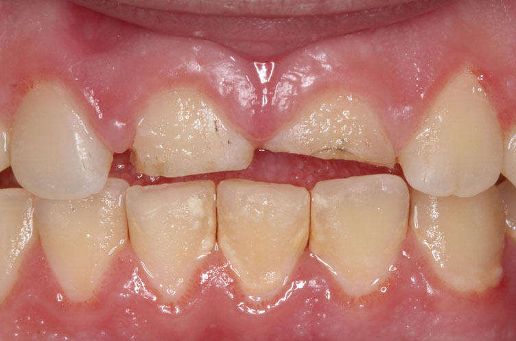
A 19-year-old Caucasian male patient attended three months after falling from a height. On examination, both maxillary central incisors were unresponsive to endo-ice and electric pulp testing. Adjacent and opposing teeth remained vital and sound. Both maxillary centrals were tender to percussion and slightly marked with color stains on the facial surface following contact with the ground during the traumatic accident. A buccal sinus tract was located between the upper right lateral and central incisor (Figure 1). Generalized plaque-induced gingivitis was noted. Grade II mobility and a 4.5 mm probing depth was associated with the deep fractured mid-lingual surfaces of the upper central incisors (Figure 2). Radiographs revealed crown-root fractures associated with the upper central incisors and external cervical resorption (Figure 3).
The patient was informed of the risks and benefits of the procedure and informed consent was gained. An immediate removable prosthesis was constructed as a back-up in case of any complications. Under local anesthesia the maxillary central incisors were caully removed with forceps via gentle rotation motions to avoid compression of the periodontal ligament (PDL) against the alveolar walls. The teeth were examined and caully rotated 180 degrees (Figure 4). Then, the upper centrals were immediately placed back into the socket adopting a more extruded coronal position (Figure 5).
The replanted teeth were stabilized with a splint for two weeks and a soft diet was advised. To prevent infection and contamination the patient was prescribed amoxicillin (500 mg every 8 hours for seven days) and chlorhexidine 0.2% rinse. After two weeks, the splint was removed using a debonding bur, and root canal treatment was initiated. No mobility was noted. A split dam was used for rubber dam isolation to prevent pressure from the clamp on the fractured teeth. The canals were cleaned with sodium hypochlorite irrigation, shaped, dried, and intracanal calcium hydroxide was applied. The patient returned after four weeks with no symptoms, no mobility and the canals were obturated with gutta-percha using a warm vertical compaction technique (Figure 6).
The next treatment stage involved restoring the upper central incisors. Electrosurgery was used around the gingival margins of both teeth which were built up using a glass fiber post and composite resin as the core (Figure 7). The teeth were then prepared for a crown (Figure 8). Lithium disilicate crowns were the choice for final restorations. Temporary crowns were removed, and the prepped teeth were cleaned. Try-in paste was used to assess the fit. Etching of the crowns was performed with 5% hydrofluoric acid to provide a micro-retentive surface. Silane Monobond Plus was applied to increase the chemical bond between the glass ceramic and silane methacrylate. Primer was applied to the bonding surface and the Emax crowns were cemented using Multilink Automix (Figure 9). The patient was followed up in six months intervals to check for periodontal health, restorative margins, esthetics, and patient symptoms. Healing of the buccal sinus tract can be seen (Figure 10). The patient was delighted that their upper front teeth were restored to achieve better function and esthetics.


Discussion
Intentional replantation with 180-degree rotation can be used in managing crown-root fractures, radicular perforations, root resorptions, or cervical caries [4]. However, this technique is contra-indicated in teeth with periodontal diseases, and divergent or dilacerated roots [5]. Intentional replantation with 180-degree rotation was perred to surgical extrusion as the rotations resulted in the subgingival lingual surface fractures to now be located more supragingival on the buccal surface. This is clinically relevant as a more coronal position of the fractured teeth can be treated more practically with greater accessibility and isolation. Furthermore, the natural dentition and interdental papilla are maintained.
The prognosis of this technique depends on the healing of the PDL. Trauma to the PDL should be avoided to achieve periodontal regeneration, cementum healing, and reduce the risk of root resorption and ankylosis. To preserve PDL viability, extraoral time was kept as short as possible with the teeth remaining out of the socket for approximately 2–3 minutes and handling of the teeth was avoided. After replantation, a coagulum forms in the PDL space which is replaced by connective tissue at 3–4 days. Following a week, the epithelium reattaches to the cementoenamel junction (CEJ) [6]. The PDL revascularizes and union of the fibers occur. The PDL healing is so advanced that after a 2-week period the periodontium establishes 2/3rd of its original adhesion, allowing for stabilization of the tooth in the alveolus.
Many replanted teeth can exhibit root resorption. Endodontic therapy after replantation reduces the risk of root resorption. Should root resorption occur this can normally be seen on radiographs within a couple of weeks and commonly occurs around the cervical 1/3rd of the root [7]. There are different types of resorptions associated with replantation. Surface resorption results from small resorptive lacunae forming in the root cementum. During replantation, damage to the cementum occurs leading to an inflammatory response as surface osteoclasts attack the root. Replacement resorption (ankylosis related) is when there is replacement of root structure by bone. This can happen following severe damage to PDL and cementum and can occur within two weeks of replanting teeth. Infection resorption relates to exposing dentinal tubules leading to a communication with an infected necrotic pulp and a subsequent inflammatory process.
Treatment options for crown-root fracture should be caully evaluated. Crown lengthening can be used to increase access by surgically inducing recession. The disadvantages of crown lengthening are challenging esthetics and time. Following crown lengthening, healing of tissues from the osseous crest averages 3 mm after three months. Although the position of the gingival margin can be established at three months, minimal changes can still occur for up to six months [8]. For this reason, it is advised to wait six months for prosthetic treatment following crown lengthening in the esthetic zone. Orthodontic extrusion involves tooth movement in a coronal direction to modify the tooth position by applying traction forces in regions of the PDL. Cons of orthodontic extrusion include a high cost and slow treatment time. Although pulp vitality can be preserved with crown lengthening and orthodontic extrusion, in this case both maxillary central incisors were non-vital on initial examination.
Intentional replantation with 180 degrees has advantages over the other treatment options discussed. Firstly, the clinical procedure takes less time compared to other options. Secondly, the fracture line can be accurately identified, isolated, and treated with greater access. This rotation technique has also been successfully demonstrated in cases where avulsed teeth are unable to be replanted back in the original position [9].
Conclusion
Intentional replantation using 180-degree rotation is a valuable treatment option for crown-root fractures. By moving the fracture line in a more coronal position through rotation and replantation, the maxillary central incisors were now deemed restorable with considerable tooth structure available and sufficient space to accommodate biological width. Greater access and isolation enabled the teeth to be treated more favorably. This technique also allows for the natural dentition and interdental papilla to be maintained. With patients having significant time constraints, intentional replantation is a viable choice of treatment. Clinical and radiographic follow-up is advised. Case selection is key with a successful outcome dependent on thorough clinical and radiographic examination, organized and sequenced treatment planning, preserving PDL health and biological healing.
REFERENCES
1.
Kaste LM, Gift HC, Bhat M, Swango PA. Prevalence of incisor trauma in persons 6-50 years of age: United States, 1988–1991. J Dent Res 1996;75 Spec No: 696–705. [CrossRef]
[Pubmed]

2.
Andreasen JO, Andreasen FM. Crown-root fractures. In: Andreasen JO, Andrasen FM, Andersson L, editor Textbook and Color Atlas of Traumatic Injuries to the Teeth. 4ed. Oxford: Blackwell; 2007. p. 314–66.
3.
Tegsjõ U, Valerius-Olsson H, Olgart K. Intra-alveolar transplantation of teeth with cervical root fractures. Swed Dent J 1978;2(3):73–82.
[Pubmed]
4.
Magini RS, Censi JC, Bianchini MA. Reimplante Intencional para Tratamento de Fissura Longitudinal: Relato clinic apos acompanhamento de um ano. Rev Bras Odontol (Impr) 1997;54(5):297–302
5.
Wolcott J, Rossman LE. Intentional replantation of endodontically treated teeth: An update. Compend Contin Educ Dent 2003;24(1):68–72, 74.
[Pubmed]
6.
Andreasen JO. A time-related study of periodontal healing and root resorption activity after replantation of mature permanent incisors in monkeys. Swed Dent J 1980;4(3):101–10.
[Pubmed]
7.
Andreasen JO, Hjorting-Hansen E. Replantation of teeth. I. Radiographic and clinical study of 110 human teeth replanted after accidental loss. Acta Odontol Scand 1966;24(3):263–86. [CrossRef]
[Pubmed]
8.
Brägger U, Lauchenauer D, Lang NP. Surgical lengthening of the clinical crown. J Clin Periodontol 1992;19(1):58–63. [CrossRef]
[Pubmed]
9.
Kondo K, Masuda I, Fukai S, Kaneko T, Horie N, Shimoyama T. Replantation of avulsed teeth using the 180-degree rotation method and a vacuum-formed splint in mixed dentition: A case report. J Oral Sci 2014;56(3):231–4. [CrossRef]
[Pubmed]
SUPPORTING INFORMATION
Author Contributions
Gurpal Hoonjan - Substantial contributions to conception and design, Drafting the article, Revising it critically for important intellectual content, Final approval of the version to be published
Maricelle Abayon - Substantial contributions to conception and design, Revising it critically for important intellectual content, Final approval of the version to be published
Data Availability StatementThe corresponding author is the guarantor of submission.
Consent For PublicationWritten informed consent was obtained from the patient for publication of this article.
Data AvailabilityAll relevant data are within the paper and its Supporting Information files.
Competing InterestsAuthors declare no conflict of interest.
Copyright© 2025 Gurpal Hoonjan et al. This article is distributed under the terms of Creative Commons Attribution License which permits unrestricted use, distribution and reproduction in any medium provided the original author(s) and original publisher are properly credited. Please see the copyright policy on the journal website for more information.