Case Report
Continuous tag and sling suture: A stable, minimally invasive technique for coronally advanced flap—A case report
1 Universidad Internacional del Ecuador, Quito, Ecuador
2 Núcleo de Odontologia – Pós-Graduação – Faculdade Arnaldo, Belo Horizonte, MG, Brazil
3 Universidad de Especialidades Espíritu Santo (UEES), Samborondón, Ecuador
4 Núcleo de Odontologia – Pós-Graduação – Faculdade Arnaldo, Belo Horizonte, MG, Brazil
Address correspondence to:
Mauricio Tinajero Aroni
Universidad Internacional del Ecuador, Jorge Fernández S/N, Quito 170411,
Equador
Access full text article on other devices

Access PDF of article on other devices
Article ID: 100052Z07MA2026
doi: 10.5348/100052Z07MA2026CR
How to cite this article
Aroni MT, Najar MG, Weisson RL, Cury VF. Continuous tag and sling suture: A stable, minimally invasive technique for coronally advanced flap—A case report. J Case Rep Images Dent 2026;12(2):1–5.ABSTRACT
Introduction: Root coverage remains a major challenge in clinical practice. The success of mucogingival surgery depends on several factors, including the choice of root coverage material and suturing technique.
Case Report: A 30-year-old male patient presented with multiple gingival recessions in the upper left quadrant. Following treatment root planing, the selected technique was a multiple coronally advanced flap combined with a connective tissue graft. During wound closure, a modification of the traditional sling and tag suturing technique was used to improve healing and clinical outcomes. Complete root coverage was achieved for all treated teeth.
Conclusion: The modified sling and tag suturing technique may be considered a less invasive and effective alternative to the original technique.
Introduction
Gingival recession is one of the most frequent mucogingival defects affecting young adults and older individuals, leading to aesthetic impairment, hypersensitivity, deficiency in biofilm control, compromising the patient’s daily life [1],[2],[3],[4]. Characterized by a multifactorial etiology, gingival recession is often linked to traumatic brushing and gingival inflammation. High frenum insertion, limited amount of keratinized tissue (KT), and thin biotype have been reported as predisposing factors for recession [4]. Smoking and systemic alterations, such as diabetes mellitus, as well as ill-fitting dental prostheses, may also contribute to the development of this defect [3],[4].
The treatment for recessions consists of root coverage surgeries, the techniques for their execution have been improved becoming less invasive and more predictable [4],[5],[6],[7]. Among the available techniques, the coronally advanced flap (CAF), with or without a subepithelial connective tissue graft (SCTG), has demonstrated satisfactory results and high rates of root coverage [4],[5],[6].
Although a predictable technique, root coverage by the CAF technique can be altered by site and patient characteristics [3],[4],[5],[6],[7]. The flap design, with minimal tension, the type of graft and choice of suture are pillars for a well-executed technique [7],[8],[9],[10]. Sutures with a uniform union of the papillae are recommended for surgeries of the CAF technique, such as sling and tag sutures [11],[12]. It is indicated because the suture is transfixed around the papilla and fixed with a knot, thus allowing greater stability of the flap [11],[12]. In addition to maintaining flap stability, the suture must allow the blood clot to remain immobile, theore, the choice of adequate suture is so important during mucogingival surgeries. Tavelli et al. [10] demonstrated in cadaver studies that marginal flap stability is best achieved in CAFs using the sling and tag suture [10],[11],[12],[13].
It has also been shown that tissue perforation by sutures reduces flap vascularity and induces a stronger postoperative inflammatory response [14],[15]. Consequently, achieving optimal surgical results necessitates appropriate flap closure and adaptation through suturing techniques that minimize tissue damage [7],[8],[9],[10],[14],[15],[16]. Modifications in suturing techniques that promote greater flap stability and less tissue trauma are desirable to optimize clinical outcomes and improve patient experience.
This manuscript describes a modification of the conventional sling and tag suturing technique, proposed as an optimal approach for achieving superior flap stability, enhanced vascularity, and minimal invasiveness in papilla suturing during CAF procedures.
Case Report
This case report is in accordance with CARE guidelines. A 30-year-old male patient, systemically healthy and non-smoker, sought dental care with a complaint of root hypersensitivity and unsatisfactory esthetics in the region of teeth 22, 23, and 24.
During the clinical examination, it was possible to observe multiple adjacent gingival recessions. The recessions were classified as Type 1 (RT1), with a detectable cementoenamel junction (CEJ) and absence of a detectable root step in all affected teeth. The recession depth was measured as the distance from the gingival margin to the CEJ using a periodontal probe. The recession depth was 3 mm for tooth 22 and 2 mm for teeth 23 and 24. All teeth with recession presented 2 mm of KT below the recessions.
The patient was informed in writing about the surgical procedure and the patient’s consent was obtained by his signature on the document.
Prior to the surgical procedure, the patient received oral hygiene instructions, supragingival scaling, and polishing. Recession depth was assessed before and after treatment. All measurements and procedures were performed by the same operator (GN). Gingival recession depth assessment was obtained using a UNC-15 periodontal probe (Hu-Friedy, Chicago, IL), with measurements rounded to the nearest millimeter. The sutures used were 5-0 poliamide monofilament (Nylon Blue NB/Poliamida Monofilamentada Azul USP 5-0, R 15 mm 3/8 Techsuture, Brazil). The width of KT was measured in millimeters using a UNC-15 periodontal probe as the distance from the free gingival margin to the mucogingival junction. The mucogingival junction was identified by the color difference and confirmed using the roll technique. The clinical attachment level (CAL) at the buccal aspect was 5 mm for tooth 22 and 4 mm for teeth 23 and 24.
Surgical Procedure
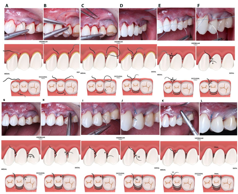
Recession coverage was performed following the technique described by Zucchelli, with a modification in the sling and tag suture, as detailed in the legend of Figure 1. A de-epithelialized connective tissue graft was used in conjunction with a coronally advanced flap. To manage postoperative discomfort, the patient was prescribed Nimesulide 100 mg every 12 hours for three days. No antibiotic therapy was administered. The patient was instructed to avoid brushing and flossing at the surgical site for three weeks. Additionally, a 0.12% chlorhexidine gluconate mouth rinse was prescribed twice daily for one week.
Follow-up was performed at one week, and sutures were removed after two weeks. Oral hygiene instructions were reinforced, and professional plaque removal was performed at each follow-up visit if necessary. A final evaluation of the clinical outcomes was conducted seven months postoperatively.
Suture Technique Description
The technique described herein is a modification of the conventional sling and tag suture. Initially, the needle is inserted buccally at the base of the mesial (or distal) surgical papilla, traversing the anatomical papilla and exiting at the palatal (or lingual) side (Figure 1A). The needle then passes around the tooth, penetrating the distal palatal papilla, and exits at the vestibular portion of the de-epithelialized anatomical papilla, re-entering at the internal base of the surgical papilla and exiting again at its vestibular side (Figure 1B).
Subsequently, the needle is inserted at the apex of the surgical papilla, transfixing the apex of the anatomical papilla and exiting at the palatal side (Figure 1C). It then returns to the vertex of the mesial palatal papilla, exits at the vestibular vertex of the de-epithelialized anatomical papilla, and penetrates internally at the vertex of the surgical papilla before exiting at the vestibular side (Figure 1D).
At this stage, a first double knot can be executed to secure the suture position (Figure 1E). The needle is then passed back through the interproximal space without engaging the papillae, in a palatal direction, wrapping around the tooth, re-entering the embrasure, and emerging buccally (Figure 1F and Figure 1G). Subsequently, the needle is inserted into the loop formed by the suspension suture (Figure 1H), passed back through the embrasure from the palatal to the mesial side, and emerges buccally, where the second simple semi-knot for containment is tied (Figure 1K). The suture is completed with a third simple security knot (Figure 1L).
Follow-up was performed at one week, and sutures were removed after two weeks. Oral hygiene instructions were reinforced, and professional plaque removal was performed at each follow-up visit if necessary. A final evaluation of the clinical outcomes was conducted seven months postoperatively.
Healing was uneventful, with minimal self-reported discomfort. The patient was followed up for seven months. Complete root coverage was achieved for all treated teeth.
No adverse events were observed during the follow-up period. The buccal CAL was reduced to 3 mm for tooth 22 and 2 mm for teeth 23 and 24, corresponding to a clinical attachment gain of 2 mm for all treated teeth. The patient reported the elimination of dental hypersensitivity, and high satisfaction with the gingival tissue contour, texture, color, and esthetics in general.
Discussion
The clinical course presented in this case report is consistent with the preoperative classification of gingival recession, as diagnosed. Complete root coverage was achieved, a result consistent with expectations when utilizing the coronally advanced flap procedure in combination with a connective tissue graft, as previously documented [2],[3],[4],[5],[6]. Subtle changes in suturing can provide benefits to the patient, as reducing the number of papillary perforations results in less tissue trauma, reduced inflammation, and improved healing. In addition, this suture can reduce the surgical time, which generates greater comfort for the patient and the dental surgeon, who optimizes their time.
The modified suturing technique proposed in this study demonstrates the capability to attain clinical results that are comparable to those established in the literature but with a simplification of the performance of the best-performing suture in cadaver studies [10]. The result of gingival recession coverage can be impacted by the choice and execution of sutures.
Sutures contribute to predictable root coverage by ensuring proper flap stabilization in the correct position and intimate adaptation of the wound margins to the root surface. This minimizes micromovements, stabilizes the blood clot, and promotes optimal wound sealing [17],[18],[19],[20]. Additionally, achieving tension-free wound closure is essential to prevent suture or tissue breakdown, marginal necrosis of the flap resulting in early and late wound dehiscence [7],[8],[9],[19].
While simple sutures are easier to perform during CAF closure, they have been shown to be less effective in flap stabilization. The sling and tag technique has demonstrated superior outcomes in terms of CAF stability in both single and multiple gingival recession coverage techniques [10]. The modified technique described here combines the sling and tag suture with a suspender technique, reducing trauma to the flap tissues while maintaining coaptation, as it requires one less needle perforation per papilla and is more time-efficient compared to the traditional tag and sling suture [13]. Thus, this technique may be theoretically indicated for mucogingival procedures involving both multiple and single gingival recessions.
Various methods for stabilizing gingival flaps have been proposed, but clinical comparisons of these techniques are rare. Yadav et al. [14] described a tripod stabilization technique for SCTG and CAF in isolated gingival recession, reporting high patient esthetic satisfaction, a high rate of root coverage, and limited postoperative morbidity [14]. Chacón Ramírez et al. [17], described a “V-Reverse” suture technique that also enhances SCTG and CAF stabilization during root coverage procedures and papilla augmentation [17]. Rebele et al. [21] introduced the vertical double-crossed suture, which aids in graft and flap stabilization and coronal positioning by securing sutures in an adjacent composite material placed in the interproximal space of the treated teeth [21]. Other stabilization methods have been tested and shown positive results when combined with suture techniques. For instance, surgical cyanoacrylate has improved the stability of tag and sling sutures in ex vivo flaps (Pabst et al. [18]), although its instability and toxicity have not been tested for in vivo applications [18]. All the aforementioned techniques share the goal of improving vascular stability without compromising flap vascularization. A notable advantage of the proposed technique is its simplified execution, as it does not require composite or adhesive fixation points.
It is expected that new studies will be carried out comparing the proposed suture with others already existing, evaluating patient esthetic satisfaction and clinical outcomes.
Conclusion
The proposed technique can be considered satisfactory for promoting a tension-free flap closure when used combined with CAF for recession coverage. The simplicity in the execution of the technique makes it attractive, as it reduces clinical execution time, making it beneficial to both the patient and the professional. Theore, the technique is a valuable addition to the sutures already used in mucogingival surgeries.
REFERENCES
1.
Imber JC, Kasaj A. Treatment of gingival recession: When and how? Int Dent J 2021;71(3):178–87. [CrossRef]
[Pubmed]

2.
Cortellini P, Bissada NF. Mucogingival conditions in the natural dentition: Narrative review, case definitions, and diagnostic considerations. J Periodontol 2018;89 Suppl 1:S204–13. [CrossRef]
[Pubmed]
3.
Yadav VS, Gumber B, Makker K, Gupta V, Tewari N, Khanduja P, et al. Global prevalence of gingival recession: A systematic review and meta-analysis. Oral Dis 2023;29(8):2993–3002. [CrossRef]
[Pubmed]
4.
Chambrone L, Pini Prato GP. Clinical insights about the evolution of root coverage procedures: The flap, the graft, and the surgery. J Periodontol 2019;90(1):9–15. [CrossRef]
[Pubmed]
5.
Tavelli L, Barootchi S, Nguyen TVN, Tattan M, Ravidà A, Wang HL. Efficacy of tunnel technique in the treatment of localized and multiple gingival recessions: A systematic review and meta-analysis. J Periodontol 2018;89(9):1075–90. [CrossRef]
[Pubmed]
6.
Chambrone L, Botelho J, Machado V, Mascarenhas P, Mendes JJ, Avila-Ortiz G. Does the subepithelial connective tissue graft in conjunction with a coronally advanced flap remain as the gold standard therapy for the treatment of single gingival recession defects? A systematic review and network meta-analysis. J Periodontol 2022;93(9):1336–52. [CrossRef]
[Pubmed]
7.
Zuhr O, Rebele SF, Cheung SL, Hürzeler MB; Research Group on Oral Soft Tissue Biology and Wound Healing. Surgery without papilla incision: Tunneling flap procedures in plastic periodontal and implant surgery. Periodontol 2000 2018;77(1):123–49. [CrossRef]
[Pubmed]
8.
Burkhardt R, Preiss A, Joss A, Lang NP. Influence of suture tension to the tearing characteristics of the soft tissues: An in vitro experiment. Clin Oral Implants Res 2008;19(3):314–9. [CrossRef]
[Pubmed]
9.
Burkhardt R, Lang NP. Role of flap tension in primary wound closure of mucoperiosteal flaps: A prospective cohort study. Clin Oral Implants Res 2010;21(1):50–4. [CrossRef]
[Pubmed]
10.
Tavelli L, Barootchi S, Ravidà A, Suárez-López Del Amo F, Rasperini G, Wang HL. Influence of suturing technique on marginal flap stability following coronally advanced flap: A cadaver study. Clin Oral Investig 2019;23(4):1641–51. [CrossRef]
[Pubmed]
11.
de Sanctis M, Zucchelli G. Coronally advanced flap: A modified surgical approach for isolated recession-type defects: Three-year results. J Clin Periodontol 2007;34(3):262–8. [CrossRef]
[Pubmed]
12.
Zucchelli G, De Sanctis M. The coronally advanced flap for the treatment of multiple recession defects: A modified surgical approach for the upper anterior teeth. J Int Acad Periodontol 2007;9(3):96–103.
[Pubmed]
13.
Huang LH, Wang HL. Sling and tag suturing technique for coronally advanced flap. Int J Periodontics Restorative Dent 2007;27(4):379–85.
[Pubmed]
14.
Yadav VS, Singh N, Bhatia A, Kamra P, Yadav R. A modified suturing protocol for tripod stabilization of connective tissue graft and coronal advancement of tunnel flap for treatment of isolated gingival recession. Int J Periodontics Restorative Dent 2022;42(1):e9–14. [CrossRef]
[Pubmed]
15.
Javed F, Al-Askar M, Almas K, Romanos GE, Al-Hezaimi K. Tissue reactions to various suture materials used in oral surgical interventions. ISRN Dent 2012;2012:762095. [CrossRef]
[Pubmed]
16.
Cairo F, Nieri M, Cincinelli S, Mervelt J, Pagliaro U. The interproximal clinical attachment level to classify gingival recessions and predict root coverage outcomes: An explorative and reliability study. J Clin Periodontol 2011;38(7):661–6. [CrossRef]
[Pubmed]
17.
Chacón Ramírez GJ, Tavelli L, Barootchi S, Wang HL. “V-reverse” suturing technique for tunnel soft tissue graft and flap stabilization: Technique illustration. Clin Adv Periodontics 2021;11(3):129–33. [CrossRef]
[Pubmed]
18.
Pabst A, Becker P, Kuchen R, Schumann S, Kasaj A. A comparative study of cyanoacrylate-based tissue adhesive and surgical sutures on marginal flap stability following coronally advanced flap. Clin Oral Investig 2023;28(1):5. [CrossRef]
[Pubmed]
19.
Wang M, Zhan Y, Liu F. Modified suture technique for stabilization of connective tissue graft in immediate implant placement and provisionalization: A short technical report. Int J Periodontics Restorative Dent 2023;43(1):e11–8. [CrossRef]
[Pubmed]
20.
Yang T, Cao Y, Zhang X. Update on suspensory suture techniques in reconstructive periodontal surgeries. Oral Maxillofac Surg 2024;28(2):539–45. [CrossRef]
[Pubmed]
21.
Zuhr O, Rebele SF, Thalmair T, Fickl S, Hürzeler MB. A modified suture technique for plastic periodontal and implant surgery – The double-crossed suture. Eur J Esthet Dent 2009;4(4):338–47.
[Pubmed]
SUPPORTING INFORMATION
Author Contributions
Mauricio Tinajero Aroni - Substantial contributions to conception and design, Analysis of data, Interpretation of data, Drafting the article, Revising it critically for important intellectual content, Final approval of the version to be published
Maria das Graças Najar - Acquisition of data, Analysis of data, Drafting the article, Final approval of the version to be published
Roberto Ledergerber Weisson - Analysis of data, Interpretation of data, Revising it critically for important intellectual content, Final approval of the version to be published
Vanessa Frazao Cury - Acquisition of data, Analysis of data, Revising it critically for important intellectual content, Final approval of the version to be published
Data Availability StatementThe corresponding author is the guarantor of submission.
Consent For PublicationWritten informed consent was obtained from the patient for publication of this article.
Data AvailabilityAll relevant data are within the paper and its Supporting Information files.
Competing InterestsAuthors declare no conflict of interest.
Copyright© 2026 Mauricio Tinajero Aroni et al. This article is distributed under the terms of Creative Commons Attribution License which permits unrestricted use, distribution and reproduction in any medium provided the original author(s) and original publisher are properly credited. Please see the copyright policy on the journal website for more information.