| Table of Contents | |
|
Case Report
| ||||||
| Mucosal defect repair with a polyglycolic acid sheet and fibrin glue after resection of large pleomorphic adenoma of the palate | ||||||
| Harusachi Kanazawa1, Atsushi Kasamatsu2, Katsuhiro Uzawa3 | ||||||
|
1DDS, PhD, Chief, Division of Dentistry and Oral-Maxillofacial Surgery, Sanmu Medical Center, 167 Naruto Sanmu City, Chiba 289-1326, Japan.
2DDS, PhD, Instructor, Department of Dentistry and Oral-Maxillofacial Surgery, Chiba University Hospital, 1-8-1 Inohana, Chuo-ku, Chiba City, Chiba 260-8670, Japan. 3DDS, PhD, Associate professor, Department of Dentistry and Oral-Maxillofacial Surgery, Chiba University Hospital, 1-8-1 Inohana, Chuo-ku, Chiba City, Chiba 260-8670, Japan. | ||||||
| ||||||
|
[HTML Abstract]
[PDF Full Text]
[Print This Article]
[Similar article in Pumed] [Similar article in Google Scholar] |
| How to cite this article: |
| Kanazawa H, Kasamatsu A, Uzawa K. Mucosal defect repair with a polyglycolic acid sheet and fibrin glue after resection of large pleomorphic adenoma of the palate. J Case Rep Images Dent 2016;2:33–36. |
|
Abstract
|
|
Introduction:
Treatment of choice for pleomorphic adenoma of the hard palate is wide local resection of the tumor including the overlying mucosa and underlying periosteum. Most conventional methods for covering the resulting palatal wound are prosthetic devices. In this case, we examined the validity of grafting a polyglycolic acid sheet and fibrin glue over a mucosal defect of the palate with a bony surface, as a substitute for a surgical splint.
Case Report: A 39-year-old male presented with a large, solid mass located on the right hard palate. Fine-needle aspiration cytology suggested pleomorphic adenoma. The patient underwent wide local resection of the lesion, and the mucosal defect was immediately covered with polyglycolic acid sheets, that were fixed with a fibrin glue spray. These sheets were tightly placed and remained in the wound, which led to complete epithelialization of the wound surface. Conclusion: Grafting the polyglycolic acid sheet with fibrin glue fixation is a useful substitute for the conventional surgical splint to cover a mucosal defect of the hard palate. | |
|
Keywords:
Mucosal defect, Palate, Pleomorphic adenoma, Polyglycolic acid sheet
| |
|
Introduction
| ||||||
|
Pleomorphic adenoma (PA) of the hard palate is typically located on the posterior third, lateral to the midline. Pleomorphic adenoma at minor salivary gland sites (e.g., the upper lip or buccal mucosa) are mobile and covered by normal mucosa, whereas lesions of the hard palate are restrained by mucosa that is tightly bound to palatal bone, and tend to lack a well-defined capsule [1]. Therefore, the treatment of choice for PA of the hard palate is wide local resection of the tumor including the overlying mucosa and underlying periosteum. Most traditional and conventional methods for covering the resulting open palatal wound are prosthetic devices (e.g., a prefabricated acrylic surgical splint) [2] [3]. Polyglycolic acid (PGA) sheets, a soft non-woven fabric, followed by its fixation using fibrin glue spray has been used to cover wounds and to prevent bleeding and leakage during surgery on the liver, pancreas, and lung because of its ability to be strongly affixed to the wound [4] [5]. We, therefore, examined the validity of grafting these sheets over a large mucosal defect of the hard palate with a bony surface, as a substitute for a surgical acrylic splint. | ||||||
|
Case Report
| ||||||
|
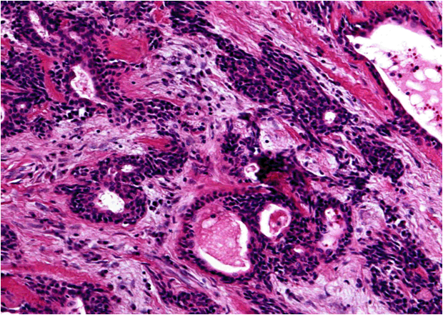
A 39-year-old Japanese man presented to our clinic with a chief complaint of having difficulty swallowing because of a painless mass on the palate. The patient reported that the lesion had slowly increased in size over the last six years. There was no relevant past medical history, and his general examination was normal. Intraoral examination revealed a broad-based, round mass with normal mucosa measuring 5.3×4.3×3.6 cm predominantly on the right side of the hard palate. It also extended laterally to the right alveolar ridge, crossing the midline (Figure 1). Computed tomography (CT) scan revealed a well-defined, expansive bone resorption of the adjacent right upper alveolar ridge of the molar region (Figure 2A). Magnetic resonance imaging (MRI) scan revealed a well-circumscribed mass with hyperintensity area on short T1 inversion recovery (STIR) or T2-weighted images (Figure 2B). No invasion of tissues adjacent to the palate was observed. Fine-needle aspiration cytology showed clusters of piled-up polygonal epithelial cells and spindle-shaped mesenchymal cells but no atypical cells, suggesting PA. Wide local resection of the lesion was undertaken that included the overlying mucosa and underlying periosteum along with a 5-mm margin at the periphery. There was no communication with the maxillary sinus or nasal cavity. For coverage of the large mucosal defect of the hard and soft palate with surface bone exposed, we used a bio-absorbable fabric composed of PGA (Neoveil;R, Gunze, Osaka, Japan) (Figure 3A) and fibrin glue (Bolheal;R, Chemo-Sero-Therapeutic Research Institute, Kumamoto, Japan). After hemostatic treatment was completed, we covered the wound by grafting PGA sheets with a thickness of 0.15 mm on it followed by fixation with the fibrin glue spray (Figure 3B) [6] [7]. For two days after surgery, the patient was nourished via nasoenteric feeding to prevent the PGA sheets from separating before starting oral feeding. He was discharged from the hospital seven days postoperatively with the PGA sheets still well attached to the wound even after oral feeding (Figure 3B). About two weeks after surgery, the sheets started to separate gradually from the surrounding soft tissue of the wound as epithelialization progressed. The sheets remained on the palatal wound for about four weeks after surgery. The wound was subsequently replaced completely by normal mucosa after six weeks. There was no wound contraction or recurrence at the two-year follow-up (Figure 4) . Neither the PGA sheets nor the fibrin glue caused any unfavorable events. Histopathological examination of the specimen confirmed the diagnosis of PA of minor salivary gland origin. This specimen had a fibrous capsule containing diffuse sheets of polygonal epithelial cells and ductal structures on a variable background stroma composed of myxoid, hyalinized, and chondroid areas (Figure 5). | ||||||
| ||||||
|
| ||||||
| ||||||
| ||||||
| ||||||
|
Discussion
| ||||||
|
Pleomorphic adenoma is a typically well-circumscribed, encapsulated tumor in the major salivary glands, mainly the parotid gland. Pleomorphic adenoma originating from the minor salivary glands in the oral cavity, however, frequently have only a poorly developed capsule or none at all, allowing them to spread into normal surrounding tissue [1]. Hence, if the tumors of the hard palate are simply enucleated, surgical exposure of the tumor or tumor capsule risks spillage and increases the risk of recurrence. Local recurrence of PA is a risk factor for malignant transformation over a long time. The propensity for carcinoma to arise from a PA has been reported at 1.9–23.3% [8]. These local characteristics of the lesion indicate that the treatment of choice for PA of the hard palate is wide local resection that includes the overlying mucosa and the underlying periosteum. With adequate surgical resection, PA of minor salivary glands usually has little tendency for recurrence, presenting a cure rate of more than 95% [1]. Regarding the mucosal defects after wide resection of the tumor in the hard palate, the standard method for covering the wound has been placement of a surgical acrylic splint [2] [3]. Previously fabricated splints, however, must be wired to the remaining teeth or alveolar process to hold the gauze packing in place. They are also not necessarily suitable for large, open wounds (extending to the mobile soft palate), sometimes causing swallowing difficulty. Such cases require a substitute for the surgical splint; a wound dressing that stably adheres to the palatal wound with a bony surface, and protects the wound during the period of complete epithelialization. Unfortunately, most wound-protective biomaterials–e.g., synthetic plastic, porcine xenograft, artificial skin, collagen membrane–are only a temporary cover for the surface of the wound, often detaching from the mucosal wound within a few days [6]. In the case reported herein, we used a new, biodegradable, mesh sheet composed of PGA to cover the open wound of the palate after surgery. The PGA sheets with fixation by fibrin glue have been applied to prevent bleeding and leakage following surgery of the liver, pancreas, and lung surgery [called the mucosal defect covered with fibrin glue and PGA sheets (MCFP) technique] [4] [5]. No unfavorable event caused by the PGA sheets or the fibrin glue has yet been reported. Recently, this MCFP technique was used to protect the raw surface after partial resection of early oral and mesopharyngeal cancer instead of the usual tie-over dressing with autologous split-thickness skin grafts [6][7]. It resulted in promoting early oral feeding because of rapid relief from postoperative pain, causing early epithelialization, and preventing scar contracture. Our case also revealed that the MCFP technique is available for covering large mucosal defects with underlying bone in the palate. It has excellent operability with adequate adhesive force and sealing effect for the wound. It also does not peel off from the underlying bony surface until epithelialization is complete. This simple method may become a useful substitute for the conventional surgical acrylic splint. However, complete hemostasis before placing the PGA sheets are required to prevent the sheets from prematurely peeling off the open palatal wound. | ||||||
|
Conclusion
| ||||||
|
Grafting the polyglycolic acid sheet with fibrin glue fixation is a simple operation for covering the large mucosal defect of the hard palate. These sheets were tightly placed and remained in the palatal wound, which led to complete epithelialization of the wound surface. It may become a useful substitute for the conventional surgical splint. | ||||||
|
References
| ||||||
| ||||||
|
[HTML Abstract]
[PDF Full Text]
|
|
Author Contributions
Harusachi Kanazawa – Substantial contributions to conception and design, Acquisition of data, Analysis and interpretation of data, Drafting the article, Revising it critically for important intellectual content, Final approval of the version to be published Atsushi Kasamatsu – Analysis and interpretation of data, Revising it critically for important intellectual content, Final approval of the version to be published Katsuhiro Uzawa – Analysis and interpretation of data, Revising it critically for important intellectual content, Final approval of the version to be published |
|
Guarantor of submission
The corresponding author is the guarantor of submission. |
|
Source of support
None |
|
Conflict of interest
Authors declare no conflict of interest. |
|
Copyright
© 2016 Harusachi Kanazawa et al. This article is distributed under the terms of Creative Commons Attribution License which permits unrestricted use, distribution and reproduction in any medium provided the original author(s) and original publisher are properly credited. Please see the copyright policy on the journal website for more information. |
|
|