 |
Case Report
Using iTero Element intra-oral scanner to scan for implant-supported fixed complete dentures
1 Owner/Lead Doctor at Barwon Heads Dental, 43 Hitchcock Avenue, Barwon Heads, Victoria 3227, Australia
2 Clinical Trainer at INVISALIGN Singapore PTE. LTD, 491B River Valley Road, Singapore 248373, Singapore
3 Sr. Manager Global Clinical Applications at Align Technology LTD, 3rd Ariel Sharon Boulevard, Or Yehuda 6037606, Israel
Address correspondence to:
Jack Bruce Milgate
Owner/Lead Doctor at Barwon Heads Dental, 43 Hitchcock Avenue, Barwon Heads, Victoria 3227,
Australia
Message to Corresponding Author
Article ID: 100036Z07JM2021
Access full text article on other devices

Access PDF of article on other devices


How to cite this article
Milgate JB, Sarawgi N, Zary R. Using iTero Element intra-oral scanner to scan for implant-supported fixed complete dentures. J Case Rep Images Dent 2021;7: 100036Z07JM2021.ABSTRACT
Introduction: Impressions in implant dentistry aim to accurately relate an analogue of the implant or implant abutment to the other structures in the dental arch. The impression material, impression technique, implant angulation, and the number of implants, all affect the accuracy. In the case of implant-supported fixed complete dentures (ISFCDs) traditional impression techniques require the doctor to accurately capture multiple units in one step. Doctors often encounter cases where one unit is not captured properly while another is perfect, so they take another impression only to find that they captured the problematic unit, but the impression of the previously perfect unit is no longer acceptable. Recent data suggests that intra-oral digital impressions may be considered a reliable alternative to conventional impression materials for ISFCDs. The ability to “segment” the impression process of large restorations and scan different segments individually eliminates much of the uncertainty that is part of taking a multi-unit conventional impression. It also ensures that units are scanned at the appropriate time, when in an ideal soft tissue condition.
Case Report: We report the case of a 48-year-old male presenting with a chief complaint of difficulty to eat due to mobile removable denture, looking for a fixed solution. This case report reviews in detail the steps followed to acquire an accurate digital impression for implant-supported fixed complete dentures using the iTero Element intra-oral scanner.
Conclusion: Digital review allowed for simpler design and easier transfer of occlusal records.
Keywords: Dental equipment, Dental implantation, Dental prosthesis, Mouth rehabilitation
Introduction
Obtaining accurate dental impressions for implant-supported fixed complete dentures (ISFCDs) is a challenge faced by clinicians on a daily basis. Ill-fitting prostheses may add unnecessary strain on the various prosthetic components in the system and ultimately result in complications [1],[2],[3],[4],[5].
Clinical studies assessing the threshold for clinically acceptable fit of ISFCDs have reported that it lies within a range of 59–200 μm [6],[7],[8]].
The advantages of digital scanning include the elimination of errors during the procedure, dispensing and polymerization stages of conventional impression materials. Eliminating the need for disinfection, shipping to the laboratory, and increased patient comfort also help to reduce errors [9],[10]. Digital intra-oral scanners (IOS) acquire single images that are stitched together using a “best-fit” algorithm to produce a virtual 3D model. Stitching can introduce errors into large scan distance such as the full-arch situation [11],[12],[13].
Our review found only two relevant publications from in vivo prospective trials. In 2019, Paolo et al. used clinical and radiological endpoints to compare conventional versus digital impressions in 50 edentulous patients. In this trial intraoral scanning reported to show satisfactory accuracy, providing a reliable alternative in clinical practice for implant full-arch rehabilitations [14]
More recently, Konstantinos et al. published a prospective clinical study to compare for the first time the accuracy of digital and conventional maxillary implant impressions in 16 edentulous patients. The 162 μm found as mean 3D deviation between the casts generated from full-arch digital and the conventional impression, appears to be in agreement with what has been previously reported in the literature and within the aforementioned threshold for clinically acceptable fit [15].
To the best of our knowledge this is the first case report to describe full arch implant workflow utilizing the iTero Element intra-oral scanner.
Case Report
We report the case of a 48-year-old male with a chief complaint of difficulty to eat due to mobile removable denture, looking for a fixed solution.
The patient had a history of failing dentition due to chronic periodontitis, Type II diabetes mellitus, and is a current smoker with a history of long-term smoking.
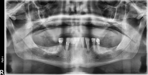
On intra-oral examination, the patient presented with an edentulous upper arch and a partially edentulous lower arch with periodontally compromised lower dentition. The patient was wearing an acrylic removable complete denture for the upper arch. The denture presented an intentional midline diastema that the patient wanted replicated in the final prosthesis. The patient was evaluated using an orthopantomogram (OPG) and a treatment plan was formulated to maintain lower failing dentition while the patient underwent an all-on-six implant supported denture for the upper jaw (Figure 1). The patient was referred to the periodontal surgeon for implant placement.
Surgical procedure for implant placement
Implant surgery was performed by periodontal surgeon based on cone-beam computed tomography (CBCT) analysis. It was a free hand surgery without the use of surgical guide. The surgery was performed keeping in consideration the patient’s medical and habitual history.
The patient had upper dentition removed nine months prior to initial implant fixture placement.
Initially 6 × Straumann BL Roxolid implants were placed. At 12 weeks integration testing, two implants showed poor osseointegration and were replaced. 10 weeks later, review of these fixtures revealed that one of the implants failed to integrate (21 area). Remaining five implants tested as stable and osseointegrated.
Taking into account the patient’s periodontal condition, medical history and continued smoking habits, it was deemed essential that a permanent restoration should be designed to allow easy maintenance and cleansing in combination with strict oral hygiene instruction and follow-ups.
Restoration for the implants
The digital method (iTero Element 2, Align Technology, Inc.) was utilized for the entire restorative process. The following steps were used to achieve the desired outcome:
- Five Straumann BL scan bodies were ordered and utilized for the restorative phase.
- Patient’s denture was scanned in the mouth to use as a copy for the final restoration (patient requested final prostheses to have the same aesthetics including midline diastema as current prostheses). Scanning the denture in situ also allowed recognition of soft tissue landmarks (frenulum) which then allowed for cross-scan calibration, articulation, and mounting of the implant scan [including replicating occlusal vertical dimension (OVD) and occlusal position].
- Lower dentition was scanned first, with the healing abutments in the upper still in place (Figure 2).
- Scanning lower prior to removal of healing abutments, minimizes time with healing abutment removed and is favorable to minimize soft tissue “collapse,” enabling capture of emergence profile and reducing discomfort to the patient.
- Upper scan was performed after taking an OPG to confirm correct seating of scan bodies (Figure 3).
- Upper scan was performed to include frenulum and full sulcus/palate (Figure 4).
Scan protocol for digital impression (iTero Element):
- The scans were performed using the scanning protocol of occlusal-palatal/lingual-buccal (Figure 5).
- It was ensured that the head of the scanner is placed deep in the sulcus area to capture it completely while using the other side to retract the soft tissues. (The size and softness of the scanner head enabled this with maximum efficiency and minimum discomfort to the patient.)
- It was important to be vigilant while scanning multiple scan-bodies to avoid a double image of multiple scan-bodies. As the scan-bodies were identical the scanner found it difficult to differentiate between them. Placing the scanner at an angle while following the basic protocol helped avoid double images.
- Good inter-scan body soft tissue capture and ensuring the scanner angle is such that it does not capture both the scan-bodies—one that is currently being scanned and the contralateral (opposing side) scan body reduces this problem.
- The scanners ability to capture the color of the scan body fixture screw enabled getting the depth of the scan body cylinder.
- If the scan does capture multiple scan-bodies overlapping one another, the scan must be deleted and started again. There was no time constraint for this case, however, for a time constraint good scan technique and practice will reduce this issue.
After the scans were captured completely and evaluated, they were sent to the lab for fabrication of the restoration.
Lab procedure
- The restorative lab utilized EXOCAD design software for designing the prosthesis. In combination with the scans sent from iTero (including bite registration, and prostheses aesthetics (denture copy) (Figure 6).
- The try-in bar, teeth and soft tissue were 3D printed using stereolithography (SLA) resin (Figure 7).
- These were then tried in the patient to confirm the fit, midline symmetry, and the prosthesis and soft tissue interface to make sure enough space for easy cleansing and maintenance. Modifications were drawn with a single use indelible marker directly on the printed try-in prosthesis (Figure 8).
- After modifications, there was a second try-in of the bar to ensure the final prosthesis had adequate notches to ensure easy cleaning (Figure 9).
- After the design was approved, milled titanium bar and polymethyl methacrylate (PMMA) teeth were used for the final prosthesis, keeping in mind the lower periodontally compromised dentition (Figure 10).
- Abutment access holes were filled with a Teflon tape spacer and finally a modified Glass-ionomer cement. Fit was good and the patient has been on regular follow-ups to ensure maintenance (Figure 11 and Figure 12).


Discussion
The accuracy for full arch dentate scans has been evaluated previously. Review of the literature showed 10 publications where iTero Element scanner was evaluated for full arch scan accuracy:
In 2019, Keul and Güth compared the accuracy of full-arch digital impressions to conventional impressions in vivo [16]. Their conclusion was that using the iTero Element intraoral scanning device resulted in the same and for single parameters even in higher accuracy than the indirect digitalization of the impression or the gypsum cast using a desktop scanner.
Two recent comparative studies used all-on-six implant models to test iTero Element versus conventional impressions as well as several other latest generation intra-oral scanners [17],[18]. While different levels of trueness and precision were found among the included IOSs, in both studies iTero was able to provide a reliable alternative to complete-arch implant impression procedures.
Five in vitro comparative studies used different edentulous and dentate models to test full arch scans accuracy [19],[20],[21],[22],[23]. Accuracy results for iTero were all within the aforementioned threshold for clinically acceptable fit.
Dutton et al. [24] and Revilla-León et al. [25] tested several scanners, including iTero Element, for the effect of common dental substrates and lighting conditions respectively on full arch scan accuracy. The new generation of scanners was deemed remarkably accurate across all substrates. For the iTero Element scanner, chair (10,000 lux) and room (1003 lux) lighting improved the trueness and precision mean values.
This case presented several advantages in comparison to conventional flow for both the dentist and the patient. With regard to the patient: 1) in conventional impression the periodontally compromised lower teeth were at risk for iatrogenic extraction, this was overcome using digital tools that do not apply any forces. 2) The patient was more comfortable with no gag. 3) The digital workflow and the eraser tool associated with the iTero scanner provided the ability for instant review of captured data preventing inconvenience due to retakes. 4) If there were any errors in the impression, that part could be recaptured and the whole scan did not have to be repeated, making the process much faster. 5) Any non-parallelism of the implant angle and under-cut that poses difficulty in removal of conventional impression can be overcome during digital. For the dentist: 1) the seamless digital workflow that involved capturing and data transfer is one of the biggest attractions associated with this treatment type. The requirement to not package and manually deliver impressions and bite registrations increased speed and delivery of lab work. 2) The process had increased patient compliance as it was not messy, and the patient did not experience gag. 3) The disposable sleeves and digital impression minimized contact with saliva and blood, providing a safer option to prevent cross infection. 4) The ability to instantly review the impression and modify it increased the efficiency of work. That said, there were a few challenges that needs to be overcome with practice and mastering the technique. 1) The limitation of this digital transfer namely presents itself regarding the labs ability to receive and efficiently interpret the digital data provided. This can be overcome with good lab training and communication. 2) During the scanning process for a full arch rehabilitation, the scan body tends to be “double-detected,” this can be overcome with angulation of the wand at 45° to ensure capturing only the required scan body.
The patient comfort is a major aspect of digital full arch scanning that makes it an attractive proposition.
Burzynski et al. compared patient acceptance and efficiency of digital intraoral scanners and alginate impressions [26]. The results of this trial showed that subjects were more comfortable, reporting less pain and dry mouth sensations with the iTero scanner than with the other methods tested. There was a significant difference in both measured time and time perception between the iTero and alginate impressions arms.
The ability to have breaks, review scans, and often not re-take impressions is very attractive for the patient. Full arch implant impressions with both open and closed tray can often be cumbersome and intrusive for the patient, and when non-parallel implants present it can create difficulties regarding post poly-vinyl siloxane (PVS) setting removal.
By far the most questionable and in this case successful part of treatment is in regard to accuracy and cross-arch stability of the multiple implant scans. The scanning technique like most aspects of dentistry takes time and practice to develop proficiency and accuracy. In this case the multiple identical scan bodies required specific scanner head angulation to limit capturing of multiple scan bodies and the scanner being able to disseminate between locations. This can be further complicated with non-parallel or in implants with minimal space inter-proximally.
Overall, the ease of scanning and comfort for the patient, communication and speed of delivery with manufacturing lab and accuracy of data make full arch implant scanning an attractive option for full arch prostheses production. Limitations regarding implant position and double capture of identical scan body data can be eliminated with good operator skill and practice.
Conclusion
The accuracy of iTero Element scanner in this case appeared flawless. The final prostheses were inserted passively with nil complications. Minor occlusal adjustments were required as this is likely due to the lack of stability of the patient’s lower arch (to be restored at a later date). Digital review allowed for simpler design and easier transfer of occlusal records.
REFERENCE
1.
Örtorp A, Torsten J. CNC-milled titanium frameworks supported by implants in the edentulous jaw: A 10- year comparative clinical study. Clin Implant Dent Relat Res 2012;14(1):88–99. [CrossRef]
[Pubmed]

2.
Papaspyridakos P, Benic GI, Hogsett VL, White GS, Lal K, Gallucci GO. Accuracy of implant casts generated with splinting and non-splinting impression techniques for edentulous patients: An optical scanning study. Clin Oral Implants Res 2012;23(6):676–81. [CrossRef]
[Pubmed]
3.
Wee AG, Aquilino SA, Schneider RL. Strategies to achieve fit in implant prosthodontics: A review of the literature. Int J Prosthodont 1999;12(2):167–78.
[Pubmed]
4.
Skalak R. Biomechanical considerations in osseointegrated prostheses. J Prosthet Dent 1983;49(6):843–8. [CrossRef]
[Pubmed]
5.
Zarb GA, Schmitt A. The longitudinal clinical effectiveness of osseointegrated dental implants: The Toronto study. Part III: Problems and complications encountered. J Prosthet Dent 1990;64(2):185–94. [CrossRef]
[Pubmed]
6.
Jemt T. In vivo measurements of precision of fit involving implant-supported prostheses in the edentulous jaw. Int J Oral Maxillofac Implants 1996;11(2):151–8.
[Pubmed]
7.
Jemt T, Book K. Prosthesis misfit and marginal bone loss in edentulous implant patients. Int J Oral Maxillofac Implants 1996;11(5):620–5.
[Pubmed]
8.
Papaspyridakos P, Benic GI, Hogsett VL, et al. Accuracy of implant casts generated with splinted and non-splinted impression techniques for edentulous patients: An optical scanning study. Clin Oral Implants Res 2012;23(6):676–81. [CrossRef]
[Pubmed]
9.
Chochlidakis KM, Papaspyridakos P, Geminiani A, Chen CJ, Feng IJ, Ercoli C. Digital versus conventional impressions for fixed prosthodontics: A systematic review and meta-analysis. J Prosthet Dent 2016;116(2):184–90. [CrossRef]
[Pubmed]
10.
Papaspyridakos P, Chen CJ, Gallucci GO, Doukoudakis A, Weber HP, Chronopoulos V. Accuracy of implant impressions for partially and completely edentulous patients: A systematic review. Int J Oral Maxillofac Implants 2014;29(4):836–45. [CrossRef]
[Pubmed]
11.
van der Meer WJ, Andriessen FS, Wismeijer D, Ren Y. Application of intra-oral dental scanners in the digital workflow of implantology. PLoS One 2012;7(8):e43312. [CrossRef]
[Pubmed]
12.
Giménez B, Özcan M, Martínez-Rus F, Pradíes G. Accuracy of a digital impression system based on parallel confocal laser technology for implants with consideration of operator experience and implant angulation and depth. Int J Oral Maxillofac Implants 2014;29(4):853–62. [CrossRef]
[Pubmed]
13.
Gimenez-Gonzalez B, Hassan B, Özcan M, Pradíes G. An in vitro study of factors influencing the performance of digital intraoral impressions operating on active wavefront sampling technology with multiple implants in the edentulous maxilla. J Prosthodont 2017;26(8):650–5. [CrossRef]
[Pubmed]
14.
Cappare P, Sannino G, Minoli M, Montemezzi P, Ferrini F. Conventional versus digital impressions for full arch screw-retained maxillary rehabilitations: A randomized clinical trial. Int J Environ Res Public Health 2019;16(5):829. [CrossRef]
[Pubmed]
15.
Chochlidakis K, Papaspyridakos P, Tsigarida A, Romeo D, Chen YW, Natto Z, Ercoli C. Digital versus conventional full-arch implant impressions: A prospective study on 16 edentulous maxillae. J Prosthodont 2020;29(4):281–6. [CrossRef]
[Pubmed]
16.
Keul C, Güth JF. Accuracy of full-arch digital impressions: An in vitro and in vivo comparison. Clin Oral Investig 2020;24(2):735–45. [CrossRef]
[Pubmed]
17.
Revilla-León M, Att W, Özcan M, Rubenstein J. Comparison of conventional, photogrammetry, and intraoral scanning accuracy of complete-arch implant impression procedures evaluated with a coordinate measuring machine. J Prosthet Dent 2021;125(3):470–8. [CrossRef]
[Pubmed]
18.
Lee KM. Comparison of two intraoral scanners based on three-dimensional surface analysis. Prog Orthod 2018;19(1):6. [CrossRef]
[Pubmed]
19.
Lee KM. Comparison of two intraoral scanners based on three-dimensional surface analysis. Prog Orthod. 2018;19(1):6. [CrossRef]
[Pubmed]
20.
Mutwalli H, Braian M, Mahmood D, Larsson C. Trueness and precision of three-dimensional digitizing intraoral devices. Int J Dent 2018;2018:5189761. [CrossRef]
[Pubmed]
21.
Braian M, Wennerberg A. Trueness and precision of 5 intraoral scanners for scanning edentulous and dentate complete-arch mandibular casts: A comparative in vitro study. J Prosthet Dent 2019;122(2):129–136.e2. [CrossRef]
[Pubmed]
22.
Kim RJ, Benic GI, Park JM. Trueness of digital intraoral impression in reproducing multiple implant position. PLoS One 2019;14(11):e0222070. [CrossRef]
[Pubmed]
23.
Iturrate M, Lizundia E, Amezua X, Solaberrieta E. A new method to measure the accuracy of intraoral scanners along the complete dental arch: A pilot study. J Adv Prosthodont 2019;11(6):331–40. [CrossRef]
[Pubmed]
24.
Dutton E, Ludlow M, Mennito A, et al. The effect different substrates have on the trueness and precision of eight different intraoral scanners. J Esthet Restor Dent 2020;32(2):204–18. [CrossRef]
[Pubmed]
25.
Revilla-León M, Jiang P, Sadeghpour M, et al. Intraoral digital scans-part 1: Influence of ambient scanning light conditions on the accuracy (trueness and precision) of different intraoral scanners. J Prosthet Dent 2020;124(3):372–8. [CrossRef]
[Pubmed]
26.
Burzynski JA, Firestone AR, Beck FM, Fields HW Jr, Deguchi T. Comparison of digital intraoral scanners and alginate impressions: Time and patient satisfaction. Am J Orthod Dentofacial Orthop 2018;153(4):534–41. [CrossRef]
[Pubmed]
SUPPORTING INFORMATION
Author Contributions
Jack Bruce Milgate - Conception of the work, Design of the work, Acquisition of data, Analysis of data, Drafting the work, Revising the work critically for important intellectual content, Final approval of the version to be published, Agree to be accountable for all aspects of the work in ensuring that questions related to the accuracy or integrity of any part of the work are appropriately investigated and resolved.
Niti Sarawgi - Conception of the work, Design of the work, Acquisition of data, Analysis of data, Drafting the work, Revising the work critically for important intellectual content, Final approval of the version to be published, Agree to be accountable for all aspects of the work in ensuring that questions related to the accuracy or integrity of any part of the work are appropriately investigated and resolved.
Raviv Zary - Conception of the work, Design of the work, Acquisition of data, Analysis of data, Drafting the work, Revising the work critically for important intellectual content, Final approval of the version to be published, Agree to be accountable for all aspects of the work in ensuring that questions related to the accuracy or integrity of any part of the work are appropriately investigated and resolved.
Guaranter of SubmissionThe corresponding author is the guarantor of submission.
Source of SupportNone
Consent StatementWritten informed consent was obtained from the patient for publication of this article.
Data AvailabilityAll relevant data are within the paper and its Supporting Information files.
Conflict of InterestAuthors declare no conflict of interest.
Copyright© 2021 Jack Bruce Milgate et al. This article is distributed under the terms of Creative Commons Attribution License which permits unrestricted use, distribution and reproduction in any medium provided the original author(s) and original publisher are properly credited. Please see the copyright policy on the journal website for more information.

