 |
Case Report
Signs of molar incisor hypomineralization before eruption of the affected tooth: Case report
1 Professor of Pediatric Clinic and Patients with Disabilities at the Catholic University of Brasilia and IESB University Center, Brasilia, Brazil
2 CEO from Fenelon, Dental Radiographic Diagnosis, Brasília, Brazil
3 Department of Morphology, Genetics, Orthodontics and Pediatric Dentistry, Araraquara School of Dentistry, São Paulo State University (Unesp), Araraquara, São Paulo, Brazil
Address correspondence to:
Lourdes Santos-Pinto
SQS 116 Bloco C, apto 108, Brasília - DF, CEP 70386030,
Brazil
Message to Corresponding Author
Article ID: 100046Z07CP2024
Access full text article on other devices

Access PDF of article on other devices


How to cite this article
Peruch CMS, Barriviera M, Barriviera FA, Santos-Pinto L. Signs of molar incisor hypomineralization before eruption of the affected tooth: Case report. J Case Rep Images Dent 2024;10(2):1–6.ABSTRACT
Introduction: Clinical manifestations of molar incisor hypomineralization (MIH) can be confused with other developmental defects of the enamel, complicating early diagnosis and appropriate treatment intervention.
Case Report: This case report demonstrated that signs suggestive of the MIH defect in first molars were possible to be identified on panoramic radiographs and 3D cone beam computed tomography (CBCT) scans, before the teeth erupted. The defect was proven with the eruption of the tooth.
Conclusion: Panoramic radiography and CBCT were radiological alternative for the early identification of signs of MIH. However, CBCT should not be routinely indicated due to its higher radiation dose.
Keywords: Cone beam computed tomography, Early diagnosis, Molar hypomineralization, Panoramic radiography
Introduction
Molar incisor hypomineralization (MIH) was the term suggested in 2001 to identify a qualitative enamel defect present in one or four first permanent molars, which can also affect the permanent incisors. Clinically, it is characterized by demarcated opacities varying in color from white to brown and, in the most severe cases, the hypomineralized enamel can easily break right after the tooth erupts, especially under the influence of masticatory forces [1].
It is estimated that the worldwide prevalence of MIH is 13.5%, with widely varying figures between countries. This variation may be due to the defect’s similarity to other enamel development defect and consequent difficulty in differential diagnosis, or due to the different methods, indices, ages, and populations of the studies [2],[3].
Currently, the main method for diagnosing MIH is clinical examination, aided by diagnostic criteria that seek to classify the degree of severity of the defect. A major challenge in diagnosis has been determining its true extent and depth. In addition to coloration, the extent and depth of the defect are important factors in the prognosis of the treatment of these teeth, since these factors influence the treatment decision by defining when we can preserve the defect and mask it or remove it and cover the affected regions with restorations [4]. It is also important to consider that the size and type of lesion are important clinical parameters for assessing the risk of posteruptive breakdown (PEB) on enamel opacities related to MIH [5].
Recently, a diagnostic tool, light transillumination, has been used preoperatively to assess opacity extension in the enamel, to confirm lesion transformation and to monitor the infiltration during the treatment [6]. However, for the complementary diagnosis of these defects, the conventional imaging method often used for dental caries, such as radiographs, offers low spatial resolution because these defects are superimposed on a large volume of tooth structure.
The limitations inherent in traditional radiography for the diagnosis of dental caries are mainly due to the 2D representation of caries lesions, which are actually 3D structures, and which can lead to the loss of valuable information. In addition, the radiographic appearance of a lesion can change dramatically depending on the projection geometry chosen. Replacing radiographic film with digital sensors has not corrected these limitations. However, cone beam computed tomography (CBCT) is a superior imaging modality to intraoral digital imaging systems and offers numerous advantages, including multi-plane reconstruction, high accuracy, and low exposure time [7],[8].
Cone beam computed tomography has shown superior sensitivity to analog or digital radiographs in identifying incipient caries lesions; however, given the higher radiation dose, its use cannot be justified as routine caries detection [9]. However, if CBCT scans are carried out for other purposes, incipient caries lesions should be reported since artifact-free CBCT images had higher diagnostic accuracy than intraoral X-ray radiography for detection of all grades of proximal caries [10].
If the initial caries lesion, which is characterized by enamel demineralization, can be identified on radiographs and CBCT scans, could MIH, an enamel defect characterized by hypomineralized and porous enamel, also be detected early on these images? The aim of this case report was to demonstrate the identification of images suggestive of the MIH defect in first molars, on panoramic radiographs and 3D cone beam CT scans, before the teeth erupted.
Case Report
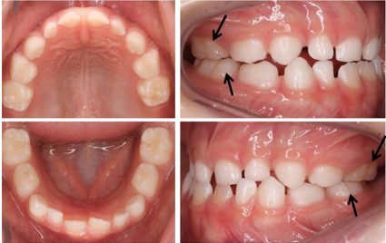
MCLB, a 5 years and 8 months boy, visited to our pediatric dentistry office and the parent’s main complaint was the demarcated opacities on posterior teeth. The patient was healthy and had no history of systemic problems. The intraoral clinical examination revealed demarcated white opacities on the buccal surface of the deciduous maxillary second molars. The pediatric dentist also diagnosed a tendency to anterior crossbite and mesial step molar relationship (Figure 1). The patient was referred to orthodontist for evaluation and treatment as early as possible.
In order to carry out the orthodontic diagnosis and treatment plan, documentation was requested, including photographs, models, teleradiography, and panoramic radiography. A computer-based analysis was performed including hard-tissue variables on cephalograms and soft-tissue variables on photographs and it was confirmed a class III skeletal discrepancies.
As the presence of opacities in deciduous teeth is a predictive factor for MIH [11], the panoramic radiograph requested by the orthodontist was used to assess the degree of tooth formation, the position, and the possible time it would take for the first permanent molars to erupt, in order to forecast and schedule return appointments. Patients with MIH have a high prevalence of caries and the affect teeth are more prone to fractures. Therefore, the patient’s return consultations at the Pediatric Dentistry office were at a reduced interval, every three months, for caries control and MIH monitoring. The patient was followed up at the orthodontist every four weeks.
In the panoramic radiograph (Axeos panoramic equipment, Dentisply–Sirona; obtained with the parameters 69 kV, 16 mA, and 14.1 seconds of rotation), it was noticed a radiolucent line at the amelodentine boundary of the occlusal surface of teeth 36 and 46 that had not yet erupted (Figure 2). Six months after the radiograph was taken, tooth 36 was present in the mouth and showed a demarcated creamy opacity on the buccal and occlusal surfaces and, in the distal portion of the central sulcus there was a discontinuity due post-eruptive fracture (Figure 3A). A therapeutic approach was taken in the area of the fracture to remove the dentin, which appeared as loose flakes, but without the characteristics of softened carious dentin (Figure 3B). The cavity was then filled with high-viscosity glass ionomer (Ketac Molar—3M) followed by the application of composite resin (Filtek Bulk Fill Flow—3M) in the remaining grooves (Figure 3C).
The patient continued with his orthodontic treatment and a CBCT (Eagle Edge CT scanner with 6×9 cm FOV, 25 second exposure, voxel size of 0.075, a focal point of 02 mm and 360° rotation) was ordered by the orthodontist to assess the treatment outcomes (Hass expander and face mask). The three-dimensional positioning visualization of the unerupted permanent teeth as well as their relationship with the teeth in the mouth and neighboring structures is important in a skeletal class III patient for a more predictable treatment plan, and more efficient patient management since in the future he may need orthognathic surgery.
At the patient’s return visit, which took place on average every three months, the CT scan requested by the orthodontist was used to assess the restored tooth (Figure 3C). The image of tooth 36 showed a radiolucent line under the enamel on the occlusal surface, similar to the radiolucent line that had been identified on the panoramic radiograph before the tooth erupted, but this tooth had dentin removed and restorative material applied due to a posteruptive fracture. However, it was noteworthy that tooth 46, which had not yet erupted, also showed a radiolucent line under the enamel on the occlusal surface, i.e., radiolucent lines similar to those seen in one year ago on the panoramic radiograph before the teeth erupted (Figure 2) were present on the CT scan and also was observed radiolucent areas in the enamel of the buccal surface (Figure 4). When tooth 46 appeared in the mouth, there was a demarcated opacity on the buccal and occlusal surfaces (Figure 5).
Analyzing the images of the upper teeth (Figure 6), radiolucent lines were present on tooth 26, which had already erupted and had a demarcated opacity and fracture (Figure 7), and on tooth 16, which had not yet erupted. When tooth 16 appeared in the mouth, there was evidence of MIH with white opacities on the buccal surface (Figure 8). It was also possible to observe white demarcated opacities on the central incisors with a small fracture on the distal edge of tooth 11. The patient was followed up by the Pediatric Dentist at intervals of three months to evaluate the first molars affected by MIH, which were protected with glass ionomer cement to avoid caries and to protect of posteruptive fracture, which is very common in affected teeth.


Discussion
Early diagnosis is important for any health problem in order to prevent it from worsening. In the case of MIH, aggravation results in post-eruptive fracture of the compromised structure and subsequent increased risk of the tooth being affected by caries disease [3].
The diagnosis of MIH is dependent on recognizing the defect during clinical examination and there are known difficulties in differential diagnosis and in determining the extent and depth of these defects. It is therefore important to look for diagnostic tools that complement the clinical examination and help us make an early diagnosis of the affected teeth and, at the same time, allow us to assess the prognosis of these teeth [12].
Radiographs have long been used as a complementary method in the diagnosis of dental caries lesions. Dentistry has also used CT scans as a complementary and auxiliary diagnostic imaging method with positive results for detecting caries lesions, including for detecting incipient caries lesions [13]. The advantage of CBCT is its accuracy in relation to the depth of the lesion, which is superior to conventional imaging tests [14].
Although CT shows good results as a complementary method for diagnosing dental caries, it is important to consider some fundamental factors when CBCT is suggested for a pediatric patient, such as the high cost and the patient’s exposure to radiation. A careful assessment is also needed to confirm that the patient can cooperate with the examination, by remaining immobile for a prolonged period [15]. This is why CT scans should only be indicated when conventional radiography does not meet the necessary diagnostic requirements. Radiation dosage is another factor to be discussed with the family, considering that diagnosis, early intervention, and frequent monitoring of teeth with MIH can help the prognosis of the affected tooth.
In the case of the patient presented here, panoramic radiography and CBCT were requested for orthodontic reasons. As the patient had hypomineralized deciduous teeth, which is a predictor of MIH [11], and the family was extremely anxious to know if the first permanent molars would be affected, we used the images to assess the patient’s permanent teeth before they erupted. It was observed that the radiolucent image present at the amelodentine boundary under the occlusal surface of the first permanent molars on the panoramic radiograph (Figure 2), which had been considered a possible artifact (some problem during the acquisition process, whether due to image processing, handling or storage), was present on the CT scan taken one year later (Figure 4). The presence of demarcated opacities was confirmed in these teeth as soon as they began to appear in the mouth (Figure 3, Figure 5, Figure 7, and Figure 8). It is noteworthy that tooth 36 already had enamel disintegration or fracture on the occlusal surface when it appeared in the mouth, reaching the dentin in the affected area, corroborating the observations of Cabral et al. (2020), that the occurrence of PEB is more frequent soon after tooth eruption [16].
In studies carried out using polarized light microscopy, no morphological changes were identified in the dentin located under the MIH defect; however, the presence of interglobular dentin was found [17]. Considering that interglobular dentin is a region of malformation and results in a hypomineralized area between the mineralized globules [18], one question remains: could this area of interglobular dentin be responsible for the radiolucent line seen on the patient’s X-ray and CT scan?
If signs of the MIH defect can be detected early by means of a good quality digital panoramic radiograph, the ideal is to take it when the first permanent molars are in Nolla stage 6 to 7, around five years of age and before their eruption. However, dental practitioners should carefully consider radiographic benefits and evidence-based indications before it is ordered. This patient started the orthodontic treatment at five years of age and panoramic radiography was part the initial records. Orthodontists find the panoramic and cephalometric radiography to be sufficient for most initial, progress, and final records [19].
In this case, CT was also an important auxiliary tool in identifying signs suggestive of MIH before the eruption of the affected tooth because CBCT offers around 512 axial slice images and produces 3D images [14]. One of the main advantages of the images obtained is the possibility of optimizing them for better viewing using navigation software, which makes it easier to identify the defect and the best image for evaluation. However, the heightened concerns about radiation risks in children restrict the justification for their use.
The early identification of MIH would help to draw up treatment plans for early intervention and prepare the family for the risks and care that must be taken to maintain the integrity of the tooth affected by MIH. In this case the early identification of these signs was used to alert the parents to the possibility of the permanent teeth being affected and to get their cooperation and commitment to frequent return visits and monitoring of the patient.
Conclusion
Based on the clinical evidence presented in this case, it is suggested that:
- On a good quality digital panoramic radiograph, signs of MIH can be observed before the eruption of the first permanent molars.
- Three-dimensional clinical techniques such as cone beam computed tomography may be a possible radiological alternative for the early identification of signs of MIH. However, CBCT should not be routinely indicated due to its higher radiation dose.
REFERENCE
1.
Weerheijm KL, Jälevik B, Alaluusua S. Molar-incisor hypomineralisation. Caries Res 2001;35(5):390–1. [CrossRef]
[Pubmed]

2.
Lopes LB, Machado V, Mascarenhas P, Mendes JJ, Botelho J. The prevalence of molar-incisor hypomineralization: A systematic review and meta-analysis. Sci Rep 2021;11(1):22405. [CrossRef]
[Pubmed]
3.
Mazur M, Corridore D, Ndokaj A, et al. MIH and dental caries in children: A systematic review and meta-analysis. Healthcare (Basel) 2023;11(12):1795. [CrossRef]
[Pubmed]
4.
Al-Azri K, Melita LN, Strange AP, et al. Optical coherence tomography use in the diagnosis of enamel defects. J Biomed Opt 2016;21(3):36004. [CrossRef]
[Pubmed]
5.
Arslanagić A, Marković N, Bajrić E, Burnazović Ristić L. Demarcated opacities as predictors of progression of the molar incisor hypomineralisation: A pilot study. Acta Stomatol Croat 2020;54(4):420–30. [CrossRef]
[Pubmed]
6.
Marouane O, Manton DJ. The use of transillumination in mapping demarcated enamel opacities in anterior teeth: A cross-sectional study. Int J Paediatr Dent 2022;32(1):49–55. [CrossRef]
[Pubmed]
7.
Esmaeilyfard R, Bonyadifard H, Paknahad M. Dental caries detection and classification in CBCT images using deep learning. Int Dent J 2024;74(2):328–34. [CrossRef]
[Pubmed]
8.
Chan EK, Wah YY, Lam WYH, Chu CH, Yu OY. Use of digital diagnostic aids for initial caries detection: A review. Dent J (Basel) 2023;11(10):232. [CrossRef]
[Pubmed]
9.
Walsh T, Macey R, Riley P, et al. Imaging modalities to inform the detection and diagnosis of early caries. Cochrane Database Syst Rev 2021;3(3):CD014545. [CrossRef]
[Pubmed]
10.
Mosavat F, Ahmadi E, Amirfarhangi S, Rafeie N. Evaluation of diagnostic accuracy of CBCT and intraoral radiography for proximal caries detection in the presence of different dental restoration materials. BMC Oral Health 2023;23(1):419. [CrossRef]
[Pubmed]
11.
Garot E, Denis A, Delbos Y, Manton D, Silva M, Rouas P. Are hypomineralised lesions on second primary molars (HSPM) a predictive sign of molar incisor hypomineralisation (MIH)? A systematic review and a meta-analysis. J Dent 2018;72:8–13. [CrossRef]
[Pubmed]
12.
Goursand D, De Souza FB, Fontes FS, Oliveira G, Lima LS, Sales NAF. Conhecendo a hipomineralização molar-incisivo: do diagnóstico ao tratamento. Brazilian Journal of Health Review 2023;6(1):1016–24. [CrossRef]
13.
Beltrán JA, León-Manco RA, Guerrero MA. Comparison of the diagnostic accuracy of cone beam computed tomography and three intraoral radiographic systems in the diagnosis of carious lesions in vitro. J Oral Res 2020;9(6):466–73. [CrossRef]
14.
Kalathingal SM, Mol A, Tyndall DA, Caplan DJ. In vitro assessment of cone beam local computed tomography for proximal caries detection. Oral Surg Oral Med Oral Pathol Oral Radiol Endod 2007;104(5):699–704. [CrossRef]
[Pubmed]
15.
Horner K, Barry S, Dave M, et al. Diagnostic efficacy of cone beam computed tomography in paediatric dentistry: A systematic review. Eur Arch Paediatr Dent 2020;21(4):407–26. [CrossRef]
[Pubmed]
16.
Cabral RN, Nyvad B, Soviero VLVM, Freitas E, Leal SC. Reliability and validity of a new classification of MIH based on severity. Clin Oral Investig 2020;24(2):727–34. [CrossRef]
[Pubmed]
17.
Fearne J, Anderson P, Davis GR. 3D X-ray microscopic study of the extent of variations in enamel density in first permanent molars with idiopathic enamel hypomineralisation. Br Dent J 2004;196(10):634–8. [CrossRef]
[Pubmed]
18.
Jayawardena C, Nandasena T, Abeywardena A, Nanayakkara D. Regional distribution of interglobular dentine in human teeth. Arch Oral Biol 2009;54(11):1016–21. [CrossRef]
[Pubmed]
19.
Abdelkarim A, Jerrold L. Clinical considerations and potential liability associated with the use of ionizing radiation in orthodontics. Am J Orthod Dentofacial Orthop 2018;154(1):15–25. [CrossRef]
[Pubmed]
SUPPORTING INFORMATION
Author Contributions
Claudia Maria de Souza Peruch - Conception of the work, Design of the work, Acquisition of data, Analysis of data, Drafting the work, Revising the work critically for important intellectual content, Final approval of the version to be published, Agree to be accountable for all aspects of the work in ensuring that questions related to the accuracy or integrity of any part of the work are appropriately investigated and resolved.
Mauricio Barriviera - Acquisition of data, Analysis of data, Revising the work critically for important intellectual content, Final approval of the version to be published, Agree to be accountable for all aspects of the work in ensuring that questions related to the accuracy or integrity of any part of the work are appropriately investigated and resolved.
Fernando Antunes Barriviera - Acquisition of data, Analysis of data, Revising the work critically for important intellectual content, Final approval of the version to be published, Agree to be accountable for all aspects of the work in ensuring that questions related to the accuracy or integrity of any part of the work are appropriately investigated and resolved.
Lourdes Santos-Pinto - Conception of the work, Design of the work, Acquisition of data, Analysis of data, Drafting the work, Revising the work critically for important intellectual content, Final approval of the version to be published, Agree to be accountable for all aspects of the work in ensuring that questions related to the accuracy or integrity of any part of the work are appropriately investigated and resolved.
Guaranter of SubmissionThe corresponding author is the guarantor of submission.
Source of SupportNone
Consent StatementWritten informed consent was obtained from the patient for publication of this article.
Data AvailabilityAll relevant data are within the paper and its Supporting Information files.
Conflict of InterestAuthors declare no conflict of interest.
Copyright© 2024 Claudia Maria de Souza Peruch et al. This article is distributed under the terms of Creative Commons Attribution License which permits unrestricted use, distribution and reproduction in any medium provided the original author(s) and original publisher are properly credited. Please see the copyright policy on the journal website for more information.

