|
Case Report
Management of peripheral ossifying fibroma removal and repair: A clinical report
1 Professsor, Department of Periodontics and Implant Dentistry, San Francisco de Quito University (USFQ), Quito, Ecuador
2 PhD Student, Department of Diagnosis and Surgery, School of Dentistry, UNESP - São Paulo State University, Araraquara, São Paulo, Brazil
3 Postdoctoral Research Associate, Department of Comprehensive Oral Health-Periodontology, University of North Carolina at Chapel Hill Adams School of Dentistry, Chapel Hill, North Carolina, USA
4 Professor, Department of Oral Pathology, School of Dentistry of Araraquara, Universidade Estadual Paulista, Araraquara, Brazil
5 Professor, Department of Diagnosis and Surgery, School of Dentistry, UNESP - São Paulo State University, Araraquara, São Paulo, Brazil
Address correspondence to:
Mauricio Andrés Tinajero Aroni
Pampite Oe4-183 y Diego de Robles, Zip code: 17-1200-841, Quito,
Ecuador
Message to Corresponding Author
Article ID: 100039Z07MA2022
Access full text article on other devices

Access PDF of article on other devices

How to cite this article
Aroni MAT, de Souza Carvalho J, Cirelli T, Spolidorio LC, Marcantonio RAC. Management of peripheral ossifying fibroma removal and repair: A clinical report. J Case Rep Images Dent 2022;8:100039Z07MA2022.ABSTRACT
Introduction: Peripheral ossifying fibroma (POF) is a gingival lesion of a reactive nature, characterized as a fibrocellular hyperplastic mass with calcified foci. It represents 2–9% of all gingival lesions, and it is the third most common lesion of all localized reactive hyperplastic lesions. The present study describes a POF removal and repair with one year of follow-up.
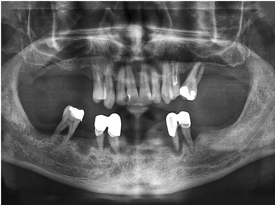
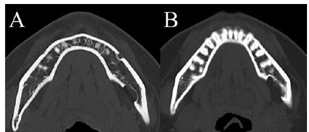
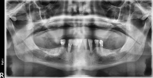
Case Report: The patient presented an increase in tissue volume in the papilla region between teeth 33 and 34, with no report of pain and with aesthetic impairment. Radiographically, no pathological changes were noted in bone and dental structures.
Conclusion: The proposed treatments were excisional biopsy associated with a subepithelial connective tissue graft (SCTG) by tunnel technique. Histologically, it revealed the presence of parakeratinized stratified squamous epithelium overlying a fibrocellular connective tissue stroma, confirming the diagnosis of peripheral ossifying fibroma. The treatment was successful, guaranteeing patient aesthetic satisfaction after one year of follow-up.
Keywords: Calcifications, Gingival hyperplasia, Ossifying fibroma
SUPPORTING INFORMATION
Author Contributions:
Mauricio Andrés Tinajero Aroni - Substantial contributions to conception and design, Acquisition of data, Analysis of data, Drafting the article, Final approval of the version to be published
Jhonatan de Souza Carvalho - Substantial contributions to conception and design, Acquisition of data, Analysis of data, Drafting the article, Final approval of the version to be published
Thamiris Cirelli - Substantial contributions to conception and design, Interpretation of data, Drafting the article, Revising it critically for important intellectual content, Final approval of the version to be published
Luis Carlos Spolidorio - Substantial contributions to conception and design, Interpretation of data, Revising it critically for important intellectual content, Final approval of the version to be published
Rosemary Adriana Chierici Marcantonio - Substantial contributions to conception and design, Interpretation of data, Revising it critically for important intellectual content, Final approval of the version to be published
Guaranter of SubmissionThe corresponding author is the guarantor of submission.
Source of SupportNone
Consent StatementWritten informed consent was obtained from the patient for publication of this article.
Data AvailabilityAll relevant data are within the paper and its Supporting Information files.
Conflict of InterestAuthors declare no conflict of interest.
Copyright© 2022 Mauricio Andrés Tinajero Aroni et al. This article is distributed under the terms of Creative Commons Attribution License which permits unrestricted use, distribution and reproduction in any medium provided the original author(s) and original publisher are properly credited. Please see the copyright policy on the journal website for more information.